 Movie
Movie Controller


+ Open data

- Basic information
| Entry | Database: PDB / ID: 5aey | ||||||
|---|---|---|---|---|---|---|---|
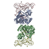
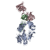
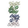
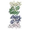
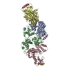
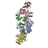
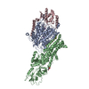
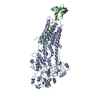
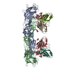
| Title | actin-like ParM protein bound to AMPPNP | ||||||
 Components Components | PLASMID SEGREGATION PROTEIN PARM | ||||||
 Keywords Keywords | STRUCTURAL PROTEIN / BACTERIAL CYTOSKELETON / PLASMID SEGREGATION / ACTIN- LIKE PROTEIN | ||||||
| Function / homology |  Function and homology information Function and homology information | ||||||
| Biological species |  | ||||||
| Method | ELECTRON MICROSCOPY / helical reconstruction / cryo EM / Resolution: 4.3 Å | ||||||
 Authors Authors | Bharat, T.A.M. / Murshudov, G.N. / Sachse, C. / Lowe, J. | ||||||
 Citation Citation | Journal: Nature / Year: 2015 Title: Structures of actin-like ParM filaments show architecture of plasmid-segregating spindles. Authors: Tanmay A M Bharat / Garib N Murshudov / Carsten Sachse / Jan Löwe /   Abstract: Active segregation of Escherichia coli low-copy-number plasmid R1 involves formation of a bipolar spindle made of left-handed double-helical actin-like ParM filaments. ParR links the filaments with ...Active segregation of Escherichia coli low-copy-number plasmid R1 involves formation of a bipolar spindle made of left-handed double-helical actin-like ParM filaments. ParR links the filaments with centromeric parC plasmid DNA, while facilitating the addition of subunits to ParM filaments. Growing ParMRC spindles push sister plasmids to the cell poles. Here, using modern electron cryomicroscopy methods, we investigate the structures and arrangements of ParM filaments in vitro and in cells, revealing at near-atomic resolution how subunits and filaments come together to produce the simplest known mitotic machinery. To understand the mechanism of dynamic instability, we determine structures of ParM filaments in different nucleotide states. The structure of filaments bound to the ATP analogue AMPPNP is determined at 4.3 Å resolution and refined. The ParM filament structure shows strong longitudinal interfaces and weaker lateral interactions. Also using electron cryomicroscopy, we reconstruct ParM doublets forming antiparallel spindles. Finally, with whole-cell electron cryotomography, we show that doublets are abundant in bacterial cells containing low-copy-number plasmids with the ParMRC locus, leading to an asynchronous model of R1 plasmid segregation. | ||||||
| History |
|
- Structure visualization
| Movie |
Movie viewer |
|---|---|
| Structure viewer | Molecule: MolmilJmol/JSmol |
- Downloads & links
-Download
| PDBx/mmCIF format | 5aey.cif.gz | 320.2 KB | Display | PDBx/mmCIF format |
|---|---|---|---|---|
| PDB format | pdb5aey.ent.gz | 261.5 KB | Display | PDB format |
| PDBx/mmJSON format | 5aey.json.gz | Tree view | PDBx/mmJSON format | |
| Others |  Other downloads Other downloads |
-Validation report
| Summary document | 5aey_validation.pdf.gz | 1 MB | Display | wwPDB validaton report |
|---|---|---|---|---|
| Full document | 5aey_full_validation.pdf.gz | 1.1 MB | Display | |
| Data in XML | 5aey_validation.xml.gz | 47.7 KB | Display | |
| Data in CIF | 5aey_validation.cif.gz | 67.1 KB | Display | |
| Arichive directory | https://data.pdbj.org/pub/pdb/validation_reports/ae/5aeyftp://data.pdbj.org/pub/pdb/validation_reports/ae/5aey | HTTPS FTP |
-Related structure data
| Related structure data |  2850MC 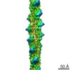 2848C 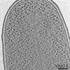 2849C  5ai7C M: map data used to model this data C: citing same article ( |
|---|---|
| Similar structure data |

-Links
 PDBj
PDBj


- Assembly
| Deposited unit | 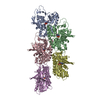
| ||||||||||||||||||||||||||||||||||||||||||||||||||||||||||||||||||||||||||||||||||||
|---|---|---|---|---|---|---|---|---|---|---|---|---|---|---|---|---|---|---|---|---|---|---|---|---|---|---|---|---|---|---|---|---|---|---|---|---|---|---|---|---|---|---|---|---|---|---|---|---|---|---|---|---|---|---|---|---|---|---|---|---|---|---|---|---|---|---|---|---|---|---|---|---|---|---|---|---|---|---|---|---|---|---|---|---|---|
| 1 |
| ||||||||||||||||||||||||||||||||||||||||||||||||||||||||||||||||||||||||||||||||||||
| Noncrystallographic symmetry (NCS) | NCS oper:
|
-Components
| #1: Protein | Mass: 35633.223 Da / Num. of mol.: 5 Source method: isolated from a genetically manipulated source Details: PARM WAS INCUBATED WITH AMPPNP / Source: (gene. exp.) #2: Chemical | ChemComp-ANP /   Mass: 506.196 Da / Num. of mol.: 5 / Source method: obtained synthetically / Formula: C10H17N6O12P3 / Comment: AMP-PNP, energy-carrying molecule analogue*YM Mass: 506.196 Da / Num. of mol.: 5 / Source method: obtained synthetically / Formula: C10H17N6O12P3 / Comment: AMP-PNP, energy-carrying molecule analogue*YM |
|---|
-Experimental details
-Experiment
| Experiment | Method: ELECTRON MICROSCOPY |
|---|---|
| EM experiment | Aggregation state: FILAMENT / 3D reconstruction method: helical reconstruction |
- Sample preparation
| Component | Name: PARM BOUND TO AMPPNP / Type: COMPLEX / Details: MICROGRAPHS COLLECTED ON AN FEI KRIOS. |
|---|---|
| Buffer solution | Name: 50 MM TRIS-HCL, 100 MM KCL, AND 1 MM MGCL2 / pH: 7 / Details: 50 MM TRIS-HCL, 100 MM KCL, AND 1 MM MGCL2 |
| Specimen | Conc.: 0.3 mg/ml / Embedding applied: NO / Shadowing applied: NO / Staining applied: NO / Vitrification applied: YES |
| Specimen support | Details: HOLEY CARBON |
| Vitrification | Instrument: FEI VITROBOT MARK IV / Cryogen name: ETHANE Details: VITRIFICATION 1 -- CRYOGEN- ETHANE, HUMIDITY- 100, INSTRUMENT- FEI VITROBOT MARK IV, |
- Electron microscopy imaging
| Experimental equipment |  Model: Titan Krios / Image courtesy: FEI Company |
|---|---|
| Microscopy | Model: FEI TITAN KRIOS / Date: Feb 2, 2014 |
| Electron gun | Electron source:  FIELD EMISSION GUN / Accelerating voltage: 300 kV / Illumination mode: FLOOD BEAM FIELD EMISSION GUN / Accelerating voltage: 300 kV / Illumination mode: FLOOD BEAM |
| Electron lens | Mode: BRIGHT FIELD / Nominal defocus max: 4000 nm / Nominal defocus min: 1000 nm / Cs: 2.7 mm |
| Image recording | Electron dose: 28 e/Å2 / Film or detector model: FEI FALCON II (4k x 4k) |
| Radiation wavelength | Relative weight: 1 |
- Processing
| 3D reconstruction | Resolution: 4.3 Å / Resolution method: FSC 0.143 CUT-OFF / Refinement type: HALF-MAPS REFINED INDEPENDENTLY / Symmetry type: HELICAL | ||||||||||||
|---|---|---|---|---|---|---|---|---|---|---|---|---|---|
| Refinement | Highest resolution: 4.3 Å | ||||||||||||
| Refinement step | Cycle: LAST / Highest resolution: 4.3 Å
|
