 Movie
Movie Controller

[English] 日本語
 Yorodumi
- PDB-5a8m: Crystal structure of the selenomethionine derivative of beta-gluc... -
+ Open data

- Basic information
| Entry | Database: PDB / ID: 5a8m | ||||||
|---|---|---|---|---|---|---|---|
| Title | Crystal structure of the selenomethionine derivative of beta-glucanase SdGluc5_26A from Saccharophagus degradans | ||||||
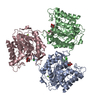
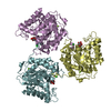
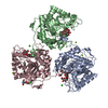
 Components Components | PUTATIVE RETAINING B-GLYCOSIDASE | ||||||
 Keywords Keywords | HYDROLASE / CAZYME / GLYCOSIDE HYDROLASE / BETA-GLUCANASE / GH5_26 | ||||||
| Function / homology |  Function and homology information Function and homology informationglucan catabolic process / hydrolase activity, hydrolyzing O-glycosyl compounds / metal ion binding Similarity search - Function | ||||||
| Biological species |  SACCHAROPHAGUS DEGRADANS 2-40 (bacteria) SACCHAROPHAGUS DEGRADANS 2-40 (bacteria) | ||||||
| Method |  X-RAY DIFFRACTION / SYNCHROTRON / SAD / Resolution: 1.86 Å X-RAY DIFFRACTION / SYNCHROTRON / SAD / Resolution: 1.86 Å | ||||||
 Authors Authors | Sulzenbacher, G. / Lafond, M. / Freyd, T. / Henrissat, B. / Coutinho, R.M. / Berrin, J.G. / Garron, M.L. | ||||||
 Citation Citation | Journal: J.Biol.Chem. / Year: 2016 Title: The Quaternary Structure of a Glycoside Hydrolase Dictates Specificity Towards Beta-Glucans Authors: Lafond, M. / Sulzenbacher, G. / Freyd, T. / Henrissat, B. / Berrin, J.G. / Garron, M.L. | ||||||
| History |
| ||||||
| Remark 700 | SHEET DETERMINATION METHOD: DSSP THE SHEETS PRESENTED AS "AB" IN EACH CHAIN ON SHEET RECORDS BELOW ... SHEET DETERMINATION METHOD: DSSP THE SHEETS PRESENTED AS "AB" IN EACH CHAIN ON SHEET RECORDS BELOW IS ACTUALLY AN 8-STRANDED BARREL THIS IS REPRESENTED BY A 9-STRANDED SHEET IN WHICH THE FIRST AND LAST STRANDS ARE IDENTICAL. SHEET DETERMINATION METHOD: DSSP THE SHEETS PRESENTED AS "BB" IN EACH CHAIN ON SHEET RECORDS BELOW IS ACTUALLY AN 8-STRANDED BARREL THIS IS REPRESENTED BY A 9-STRANDED SHEET IN WHICH THE FIRST AND LAST STRANDS ARE IDENTICAL. SHEET DETERMINATION METHOD: DSSP THE SHEETS PRESENTED AS "CB" IN EACH CHAIN ON SHEET RECORDS BELOW IS ACTUALLY AN 8-STRANDED BARREL THIS IS REPRESENTED BY A 9-STRANDED SHEET IN WHICH THE FIRST AND LAST STRANDS ARE IDENTICAL. |
- Structure visualization
| Structure viewer | Molecule: MolmilJmol/JSmol |
|---|
- Downloads & links
-Download
| PDBx/mmCIF format | 5a8m.cif.gz | 443.8 KB | Display | PDBx/mmCIF format |
|---|---|---|---|---|
| PDB format | pdb5a8m.ent.gz | 365.2 KB | Display | PDB format |
| PDBx/mmJSON format | 5a8m.json.gz | Tree view | PDBx/mmJSON format | |
| Others |  Other downloads Other downloads |
-Validation report
| Arichive directory | https://data.pdbj.org/pub/pdb/validation_reports/a8/5a8mftp://data.pdbj.org/pub/pdb/validation_reports/a8/5a8m | HTTPS FTP |
|---|
-Related structure data
| Related structure data |  5a8nC  5a8oC  5a8pC 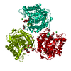 5a8qC  5a94C  5a95C C: citing same article ( |
|---|---|
| Similar structure data |

-Links
 PDBj
PDBj- Assembly
| Deposited unit | 
| |||||||||||||||||||||||||||||||||||||||||||||||||||||||||||||||||||||||||||||||||||||||||||||
|---|---|---|---|---|---|---|---|---|---|---|---|---|---|---|---|---|---|---|---|---|---|---|---|---|---|---|---|---|---|---|---|---|---|---|---|---|---|---|---|---|---|---|---|---|---|---|---|---|---|---|---|---|---|---|---|---|---|---|---|---|---|---|---|---|---|---|---|---|---|---|---|---|---|---|---|---|---|---|---|---|---|---|---|---|---|---|---|---|---|---|---|---|---|---|
| 1 |
| |||||||||||||||||||||||||||||||||||||||||||||||||||||||||||||||||||||||||||||||||||||||||||||
| Unit cell |
| |||||||||||||||||||||||||||||||||||||||||||||||||||||||||||||||||||||||||||||||||||||||||||||
| Noncrystallographic symmetry (NCS) | NCS domain:
NCS domain segments: Component-ID: _ / End auth comp-ID: LYS / End label comp-ID: LYS / Refine code: _
NCS ensembles :
NCS oper:
|
-Components
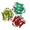
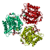
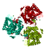
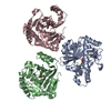
-Protein , 1 types, 3 molecules ABC
| #1: Protein | Mass: 42424.930 Da / Num. of mol.: 3 / Fragment: CATALYTIC DOMAIN, RESIDUES 1-365 / Mutation: YES Source method: isolated from a genetically manipulated source Source: (gene. exp.) SACCHAROPHAGUS DEGRADANS 2-40 (bacteria)Production host: |
|---|
-Non-polymers , 6 types, 1267 molecules 


| #2: Chemical | ChemComp-CL /  Mass: 35.453 Da / Num. of mol.: 9 / Source method: obtained synthetically / Formula: Cl Mass: 35.453 Da / Num. of mol.: 9 / Source method: obtained synthetically / Formula: Cl#3: Chemical |  Mass: 24.305 Da / Num. of mol.: 3 / Source method: obtained synthetically / Formula: Mg Mass: 24.305 Da / Num. of mol.: 3 / Source method: obtained synthetically / Formula: Mg#4: Chemical |  Mass: 96.063 Da / Num. of mol.: 3 / Source method: obtained synthetically / Formula: SO4 Mass: 96.063 Da / Num. of mol.: 3 / Source method: obtained synthetically / Formula: SO4#5: Chemical | ChemComp-GOL /  Mass: 92.094 Da / Num. of mol.: 7 / Source method: obtained synthetically / Formula: C3H8O3 Mass: 92.094 Da / Num. of mol.: 7 / Source method: obtained synthetically / Formula: C3H8O3#6: Chemical | ChemComp-PGE /  Mass: 150.173 Da / Num. of mol.: 22 / Source method: obtained synthetically / Formula: C6H14O4 Mass: 150.173 Da / Num. of mol.: 22 / Source method: obtained synthetically / Formula: C6H14O4#7: Water | ChemComp-HOH / | Mass: 18.015 Da / Num. of mol.: 1223 / Source method: isolated from a natural source / Formula: H2O |
|---|
-Details
| Has protein modification | Y |
|---|
-Experimental details
-Experiment
| Experiment | Method: X-RAY DIFFRACTION / Number of used crystals: 1 |
|---|
- Sample preparation
| Crystal | Density Matthews: 2.94 Å3/Da / Density % sol: 58.2 % / Description: NONE |
|---|---|
| Crystal grow | pH: 6.5 Details: 25% (W/V) PEG3350, 0.2 M MGCL2, 0.1 M BIS-TRIS BUFFER PH 6.5. |
-Data collection
| Diffraction | Mean temperature: 100 K |
|---|---|
| Diffraction source | Source: SYNCHROTRON / Site: SOLEIL  / Beamline: PROXIMA 1 / Wavelength: 0.979 / Beamline: PROXIMA 1 / Wavelength: 0.979 |
| Detector | Type: DECTRIS PILATUS 6M / Detector: PIXEL / Date: Mar 16, 2012 |
| Radiation | Protocol: SINGLE WAVELENGTH / Monochromatic (M) / Laue (L): M / Scattering type: x-ray |
| Radiation wavelength | Wavelength: 0.979 Å / Relative weight: 1 |
| Reflection | Resolution: 1.86→47.53 Å / Num. obs: 116426 / % possible obs: 99.4 % / Observed criterion σ(I): 0 / Redundancy: 6 % / Rmerge(I) obs: 0.1 / Net I/σ(I): 9.5 |
| Reflection shell | Resolution: 1.86→1.96 Å / Redundancy: 5.9 % / Rmerge(I) obs: 0.38 / Mean I/σ(I) obs: 3.5 / % possible all: 99.2 |
- Processing
| Software |
| ||||||||||||||||||||||||||||||||||||||||||||||||||||||||||||||||||||||||||||||||||||||||||||||||||||||||||||||||||||||||||||||||||||||||||||||||||||||||||||||||||||||||||||||||||||||
|---|---|---|---|---|---|---|---|---|---|---|---|---|---|---|---|---|---|---|---|---|---|---|---|---|---|---|---|---|---|---|---|---|---|---|---|---|---|---|---|---|---|---|---|---|---|---|---|---|---|---|---|---|---|---|---|---|---|---|---|---|---|---|---|---|---|---|---|---|---|---|---|---|---|---|---|---|---|---|---|---|---|---|---|---|---|---|---|---|---|---|---|---|---|---|---|---|---|---|---|---|---|---|---|---|---|---|---|---|---|---|---|---|---|---|---|---|---|---|---|---|---|---|---|---|---|---|---|---|---|---|---|---|---|---|---|---|---|---|---|---|---|---|---|---|---|---|---|---|---|---|---|---|---|---|---|---|---|---|---|---|---|---|---|---|---|---|---|---|---|---|---|---|---|---|---|---|---|---|---|---|---|---|---|
| Refinement | Method to determine structure: SAD Starting model: NONE Resolution: 1.86→47.33 Å / Cor.coef. Fo:Fc: 0.964 / Cor.coef. Fo:Fc free: 0.955 / SU B: 3.598 / SU ML: 0.058 / Cross valid method: THROUGHOUT / ESU R: 0.102 / ESU R Free: 0.094 / Stereochemistry target values: MAXIMUM LIKELIHOOD Details: HYDROGENS HAVE BEEN ADDED IN THE RIDING POSITIONS. U VALUES WITH TLS ADDED
| ||||||||||||||||||||||||||||||||||||||||||||||||||||||||||||||||||||||||||||||||||||||||||||||||||||||||||||||||||||||||||||||||||||||||||||||||||||||||||||||||||||||||||||||||||||||
| Solvent computation | Ion probe radii: 0.8 Å / Shrinkage radii: 0.8 Å / VDW probe radii: 1.2 Å / Solvent model: BABINET MODEL WITH MASK | ||||||||||||||||||||||||||||||||||||||||||||||||||||||||||||||||||||||||||||||||||||||||||||||||||||||||||||||||||||||||||||||||||||||||||||||||||||||||||||||||||||||||||||||||||||||
| Displacement parameters | Biso mean: 18.061 Å2
| ||||||||||||||||||||||||||||||||||||||||||||||||||||||||||||||||||||||||||||||||||||||||||||||||||||||||||||||||||||||||||||||||||||||||||||||||||||||||||||||||||||||||||||||||||||||
| Refinement step | Cycle: LAST / Resolution: 1.86→47.33 Å
| ||||||||||||||||||||||||||||||||||||||||||||||||||||||||||||||||||||||||||||||||||||||||||||||||||||||||||||||||||||||||||||||||||||||||||||||||||||||||||||||||||||||||||||||||||||||
| Refine LS restraints |
|