 Movie
Movie Controller


+ Open data

- Basic information
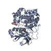
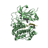
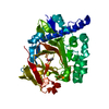
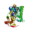
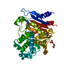
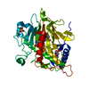
| Entry | Database: PDB / ID: 5a53 | ||||||
|---|---|---|---|---|---|---|---|
| Title | Crystal Structure of the Rpf2-Rrs1 complex | ||||||
 Components Components |
| ||||||
 Keywords Keywords | TRANSCRIPTION / 5S RNP / RIBOSOME ASSEMBLY / RRS1 / RPF2 / BRIX DOMAIN | ||||||
| Function / homology |  Function and homology information Function and homology information7S RNA binding / maturation of 5.8S rRNA from tricistronic rRNA transcript (SSU-rRNA, 5.8S rRNA, LSU-rRNA) / preribosome, large subunit precursor / ribosomal large subunit export from nucleus / endonucleolytic cleavage in ITS1 to separate SSU-rRNA from 5.8S rRNA and LSU-rRNA from tricistronic rRNA transcript (SSU-rRNA, 5.8S rRNA, LSU-rRNA) / nuclear periphery / ribosomal large subunit biogenesis / maturation of LSU-rRNA from tricistronic rRNA transcript (SSU-rRNA, 5.8S rRNA, LSU-rRNA) / assembly of large subunit precursor of preribosome / 5S rRNA binding ...7S RNA binding / maturation of 5.8S rRNA from tricistronic rRNA transcript (SSU-rRNA, 5.8S rRNA, LSU-rRNA) / preribosome, large subunit precursor / ribosomal large subunit export from nucleus / endonucleolytic cleavage in ITS1 to separate SSU-rRNA from 5.8S rRNA and LSU-rRNA from tricistronic rRNA transcript (SSU-rRNA, 5.8S rRNA, LSU-rRNA) / nuclear periphery / ribosomal large subunit biogenesis / maturation of LSU-rRNA from tricistronic rRNA transcript (SSU-rRNA, 5.8S rRNA, LSU-rRNA) / assembly of large subunit precursor of preribosome / 5S rRNA binding / ribosomal large subunit assembly / rRNA binding / nucleolus / nucleoplasm Similarity search - Function | ||||||
| Biological species |  | ||||||
| Method |  X-RAY DIFFRACTION / SYNCHROTRON / SIRAS / Resolution: 2.401 Å X-RAY DIFFRACTION / SYNCHROTRON / SIRAS / Resolution: 2.401 Å | ||||||
 Authors Authors | Madru, C. / Lebaron, S. / Blaud, M. / Delbos, L. / Rety, S. / Leulliot, N. | ||||||
 Citation Citation | Journal: Genes Dev. / Year: 2015 Title: Chaperoning 5S RNA Assembly. Authors: Madru, C. / Lebaron, S. / Blaud, M. / Delbos, L. / Pipoli, J. / Pasmant, E. / Rety, S. / Leulliot, N. | ||||||
| History |
|
- Structure visualization
| Structure viewer | Molecule: MolmilJmol/JSmol |
|---|
- Downloads & links
-Download
| PDBx/mmCIF format | 5a53.cif.gz | 76.7 KB | Display | PDBx/mmCIF format |
|---|---|---|---|---|
| PDB format | pdb5a53.ent.gz | 57.4 KB | Display | PDB format |
| PDBx/mmJSON format | 5a53.json.gz | Tree view | PDBx/mmJSON format | |
| Others |  Other downloads Other downloads |
-Validation report
| Arichive directory | https://data.pdbj.org/pub/pdb/validation_reports/a5/5a53ftp://data.pdbj.org/pub/pdb/validation_reports/a5/5a53 | HTTPS FTP |
|---|
-Related structure data
| Similar structure data |
|---|


-Links
 PDBj
PDBj- Assembly
| Deposited unit | 
| ||||||||
|---|---|---|---|---|---|---|---|---|---|
| 1 |
| ||||||||
| Unit cell |
| ||||||||
| Components on special symmetry positions |
|
-Components
| #1: Protein | Mass: 7273.322 Da / Num. of mol.: 1 / Fragment: RESIDUES 9-73 Source method: isolated from a genetically manipulated source Details: TRYPSINOLYZED SAMPLE Source: (gene. exp.) Plasmid: PET21A / Production host:  | ||||
|---|---|---|---|---|---|
| #2: Protein/peptide | Mass: 2463.886 Da / Num. of mol.: 1 / Fragment: RESIDUES 85-106 Source method: isolated from a genetically manipulated source Details: TRYPSINOLYZED SAMPLE Source: (gene. exp.) Plasmid: PET21A / Production host: | ||||
| #3: Protein | Mass: 26575.982 Da / Num. of mol.: 1 / Fragment: BRIX DOMAIN, RESIDUES 23-252 Source method: isolated from a genetically manipulated source Details: TRYPSINOLYSED SAMPLE Source: (gene. exp.) Plasmid: PET21A / Production host: | ||||
| #4: Chemical | ChemComp-SO4 /   Mass: 96.063 Da / Num. of mol.: 4 / Source method: obtained synthetically / Formula: SO4 Mass: 96.063 Da / Num. of mol.: 4 / Source method: obtained synthetically / Formula: SO4#5: Water | ChemComp-HOH / |  Mass: 18.015 Da / Num. of mol.: 93 / Source method: isolated from a natural source / Formula: H2O Mass: 18.015 Da / Num. of mol.: 93 / Source method: isolated from a natural source / Formula: H2OSequence details | PROTEOLYZE | |
-Experimental details
-Experiment
| Experiment | Method: X-RAY DIFFRACTION / Number of used crystals: 3 |
|---|
- Sample preparation
| Crystal | Density Matthews: 2.99 Å3/Da / Density % sol: 50.8 % / Description: NONE |
|---|---|
| Crystal grow | pH: 8 Details: CRYSTALS WERE OBTAINED IN 0.2M LISO4, 30 % (W/V) POLYETHYLENE GLYCOL 4000 AND 0.1 M TRIS-HCL PH 8.5, WITH A COMPLEX SOLUTION AT 15 MG/ML CONTAINING TRYPSIN. CRYSTALS WERE CRYOPROTECTED USING ...Details: CRYSTALS WERE OBTAINED IN 0.2M LISO4, 30 % (W/V) POLYETHYLENE GLYCOL 4000 AND 0.1 M TRIS-HCL PH 8.5, WITH A COMPLEX SOLUTION AT 15 MG/ML CONTAINING TRYPSIN. CRYSTALS WERE CRYOPROTECTED USING SUCCESSIVE SOAKING STEPS IN INCREASING CONCENTRATIONS OF ETHYLENE GLYCOL |
-Data collection
| Diffraction | Mean temperature: 287 K |
|---|---|
| Diffraction source | Source: SYNCHROTRON / Site: SOLEIL  / Beamline: PROXIMA 2 / Wavelength: 1.0716 / Beamline: PROXIMA 2 / Wavelength: 1.0716 |
| Detector | Type: DECTRIS PILATUS 6M / Detector: PIXEL |
| Radiation | Protocol: SINGLE WAVELENGTH / Monochromatic (M) / Laue (L): M / Scattering type: x-ray |
| Radiation wavelength | Wavelength: 1.0716 Å / Relative weight: 1 |
| Reflection | Resolution: 2.4→45.51 Å / Num. obs: 18921 / % possible obs: 99.7 % / Observed criterion σ(I): 2 / Redundancy: 11.5 % / Biso Wilson estimate: 53.99 Å2 / Rmerge(I) obs: 0.1 / Net I/σ(I): 17.82 |
| Reflection shell | Resolution: 2.4→2.49 Å / Redundancy: 11.3 % / Rmerge(I) obs: 1.09 / Mean I/σ(I) obs: 1.92 / % possible all: 97.5 |
- Processing
| Software |
| ||||||||||||||||||||||||||||||||||||||||||||||||||||||||
|---|---|---|---|---|---|---|---|---|---|---|---|---|---|---|---|---|---|---|---|---|---|---|---|---|---|---|---|---|---|---|---|---|---|---|---|---|---|---|---|---|---|---|---|---|---|---|---|---|---|---|---|---|---|---|---|---|---|
| Refinement | Method to determine structure: SIRAS Starting model: NONE Resolution: 2.401→45.505 Å / SU ML: 0.3 / σ(F): 1.36 / Phase error: 23.1 / Stereochemistry target values: ML
| ||||||||||||||||||||||||||||||||||||||||||||||||||||||||
| Solvent computation | Shrinkage radii: 0.9 Å / VDW probe radii: 1.11 Å / Solvent model: FLAT BULK SOLVENT MODEL | ||||||||||||||||||||||||||||||||||||||||||||||||||||||||
| Refinement step | Cycle: LAST / Resolution: 2.401→45.505 Å
| ||||||||||||||||||||||||||||||||||||||||||||||||||||||||
| Refine LS restraints |
| ||||||||||||||||||||||||||||||||||||||||||||||||||||||||
| LS refinement shell |
|
