 Movie
Movie Controller

[English] 日本語
 Yorodumi
- PDB-5a12: Crystal structure of Chlorite Dismutase from Magnetospirillum sp.... -
+ Open data

- Basic information
| Entry | Database: PDB / ID: 5a12 | ||||||
|---|---|---|---|---|---|---|---|
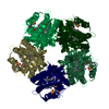
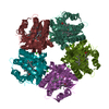
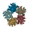
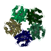
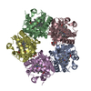
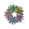
| Title | Crystal structure of Chlorite Dismutase from Magnetospirillum sp. in complex with azide | ||||||
 Components Components | CHLORITE DISMUTASE | ||||||
 Keywords Keywords | OXIDOREDUCTASE / CHLORITE / CHLORIDE / DETOXIFICATION / HEME / IRON | ||||||
| Function / homology |  Function and homology information Function and homology informationchlorite O2-lyase / chlorite O2-lyase activity / heme binding / metal ion binding Similarity search - Function | ||||||
| Biological species |  MAGNETOSPIRILLUM SP. (magnetotactic) MAGNETOSPIRILLUM SP. (magnetotactic) | ||||||
| Method |  X-RAY DIFFRACTION / SYNCHROTRON / MOLECULAR REPLACEMENT / Resolution: 1.4 Å X-RAY DIFFRACTION / SYNCHROTRON / MOLECULAR REPLACEMENT / Resolution: 1.4 Å | ||||||
 Authors Authors | Correia, H.D. / Santos-Silva, T. | ||||||
 Citation Citation | Journal: J.Phys.Chem.B / Year: 2015 Title: Ligand Binding to Chlorite Dismutase from Magnetospirillum Sp. Authors: De Schutter, A. / Correia, H. / Freire, D.M. / Rivas, M.G. / Rizzi, A.C. / Santos-Silva, T. / Gonzalez, P.J. / Van Doorslaer, S. | ||||||
| History |
| ||||||
| Remark 700 | SHEET DETERMINATION METHOD: DSSP THE SHEETS PRESENTED AS "AA" IN EACH CHAIN ON SHEET RECORDS BELOW ... SHEET DETERMINATION METHOD: DSSP THE SHEETS PRESENTED AS "AA" IN EACH CHAIN ON SHEET RECORDS BELOW IS ACTUALLY AN 8-STRANDED BARREL THIS IS REPRESENTED BY A 9-STRANDED SHEET IN WHICH THE FIRST AND LAST STRANDS ARE IDENTICAL. THE SHEETS PRESENTED AS "BA" IN EACH CHAIN ON SHEET RECORDS BELOW IS ACTUALLY AN 8-STRANDED BARREL THIS IS REPRESENTED BY A 9-STRANDED SHEET IN WHICH THE FIRST AND LAST STRANDS ARE IDENTICAL. THE SHEETS PRESENTED AS "CA" IN EACH CHAIN ON SHEET RECORDS BELOW IS ACTUALLY AN 8-STRANDED BARREL THIS IS REPRESENTED BY A 9-STRANDED SHEET IN WHICH THE FIRST AND LAST STRANDS ARE IDENTICAL. THE SHEETS PRESENTED AS "DA" IN EACH CHAIN ON SHEET RECORDS BELOW IS ACTUALLY AN 8-STRANDED BARREL THIS IS REPRESENTED BY A 9-STRANDED SHEET IN WHICH THE FIRST AND LAST STRANDS ARE IDENTICAL. THE SHEETS PRESENTED AS "EA" IN EACH CHAIN ON SHEET RECORDS BELOW IS ACTUALLY AN 8-STRANDED BARREL THIS IS REPRESENTED BY A 9-STRANDED SHEET IN WHICH THE FIRST AND LAST STRANDS ARE IDENTICAL. |
- Structure visualization
| Structure viewer | Molecule: MolmilJmol/JSmol |
|---|
- Downloads & links
-Download
| PDBx/mmCIF format | 5a12.cif.gz | 564.2 KB | Display | PDBx/mmCIF format |
|---|---|---|---|---|
| PDB format | pdb5a12.ent.gz | 467.1 KB | Display | PDB format |
| PDBx/mmJSON format | 5a12.json.gz | Tree view | PDBx/mmJSON format | |
| Others |  Other downloads Other downloads |
-Validation report
| Arichive directory | https://data.pdbj.org/pub/pdb/validation_reports/a1/5a12ftp://data.pdbj.org/pub/pdb/validation_reports/a1/5a12 | HTTPS FTP |
|---|
-Related structure data
| Related structure data |  5a13C  3q08S C: citing same article ( S: Starting model for refinement |
|---|---|
| Similar structure data |


-Links
 PDBj
PDBj- Assembly
| Deposited unit | 
| ||||||||
|---|---|---|---|---|---|---|---|---|---|
| 1 |
| ||||||||
| Unit cell |
|
-Components
| #1: Protein | Mass: 26806.664 Da / Num. of mol.: 5 / Source method: isolated from a natural source / Source: (natural) MAGNETOSPIRILLUM SP. (magnetotactic) / References: UniProt: A0A0M3KL46*PLUS, chlorite O2-lyase#2: Chemical | ChemComp-HEM /   Mass: 616.487 Da / Num. of mol.: 5 / Source method: obtained synthetically / Formula: C34H32FeN4O4 Mass: 616.487 Da / Num. of mol.: 5 / Source method: obtained synthetically / Formula: C34H32FeN4O4#3: Chemical | ChemComp-AZI /   Mass: 42.020 Da / Num. of mol.: 5 / Source method: obtained synthetically / Formula: N3 Mass: 42.020 Da / Num. of mol.: 5 / Source method: obtained synthetically / Formula: N3#4: Chemical | ChemComp-GOL /   Mass: 92.094 Da / Num. of mol.: 8 / Source method: obtained synthetically / Formula: C3H8O3 Mass: 92.094 Da / Num. of mol.: 8 / Source method: obtained synthetically / Formula: C3H8O3#5: Water | ChemComp-HOH / |  Mass: 18.015 Da / Num. of mol.: 1745 / Source method: isolated from a natural source / Formula: H2O Mass: 18.015 Da / Num. of mol.: 1745 / Source method: isolated from a natural source / Formula: H2O |
|---|
-Experimental details
-Experiment
| Experiment | Method: X-RAY DIFFRACTION / Number of used crystals: 1 |
|---|
- Sample preparation
| Crystal | Density Matthews: 2.65 Å3/Da / Density % sol: 53.7 % / Description: NONE |
|---|---|
| Crystal grow | pH: 6 / Details: 16% PEG 6000, 0.01M SODIUM CITRATE |
-Data collection
| Diffraction | Mean temperature: 77 K |
|---|---|
| Diffraction source | Source: SYNCHROTRON / Site: SLS  / Beamline: X06SA / Wavelength: 1 / Beamline: X06SA / Wavelength: 1 |
| Detector | Type: DECTRIS PILATUS / Detector: PIXEL / Date: Dec 12, 2013 |
| Radiation | Protocol: SINGLE WAVELENGTH / Monochromatic (M) / Laue (L): M / Scattering type: x-ray |
| Radiation wavelength | Wavelength: 1 Å / Relative weight: 1 |
| Reflection | Resolution: 1.4→48.84 Å / Num. obs: 268333 / % possible obs: 98.4 % / Observed criterion σ(I): 1.4 / Redundancy: 3.8 % / Rmerge(I) obs: 0.02 / Net I/σ(I): 11.4 |
| Reflection shell | Resolution: 1.4→1.42 Å / Redundancy: 3.4 % / Rmerge(I) obs: 0.74 / Mean I/σ(I) obs: 1.4 / % possible all: 94.2 |
- Processing
| Software |
| ||||||||||||||||||||||||||||||||||||||||||||||||||||||||||||||||||||||||||||||||||||||||||||||||||||||||||||||||||||||||||||||||||||||||||||||||||||||||||||||||||||||||||||||||||||||
|---|---|---|---|---|---|---|---|---|---|---|---|---|---|---|---|---|---|---|---|---|---|---|---|---|---|---|---|---|---|---|---|---|---|---|---|---|---|---|---|---|---|---|---|---|---|---|---|---|---|---|---|---|---|---|---|---|---|---|---|---|---|---|---|---|---|---|---|---|---|---|---|---|---|---|---|---|---|---|---|---|---|---|---|---|---|---|---|---|---|---|---|---|---|---|---|---|---|---|---|---|---|---|---|---|---|---|---|---|---|---|---|---|---|---|---|---|---|---|---|---|---|---|---|---|---|---|---|---|---|---|---|---|---|---|---|---|---|---|---|---|---|---|---|---|---|---|---|---|---|---|---|---|---|---|---|---|---|---|---|---|---|---|---|---|---|---|---|---|---|---|---|---|---|---|---|---|---|---|---|---|---|---|---|
| Refinement | Method to determine structure: MOLECULAR REPLACEMENT Starting model: PDB ENTRY 3Q08 Resolution: 1.4→71.44 Å / Cor.coef. Fo:Fc: 0.984 / Cor.coef. Fo:Fc free: 0.977 / SU B: 2.076 / SU ML: 0.035 / Cross valid method: THROUGHOUT / ESU R: 0.047 / ESU R Free: 0.047 / Stereochemistry target values: MAXIMUM LIKELIHOOD Details: HYDROGENS HAVE BEEN ADDED IN THE RIDING POSITIONS. U VALUES REFINED INDIVIDUALLY
| ||||||||||||||||||||||||||||||||||||||||||||||||||||||||||||||||||||||||||||||||||||||||||||||||||||||||||||||||||||||||||||||||||||||||||||||||||||||||||||||||||||||||||||||||||||||
| Solvent computation | Ion probe radii: 0.8 Å / Shrinkage radii: 0.8 Å / VDW probe radii: 1.2 Å / Solvent model: MASK | ||||||||||||||||||||||||||||||||||||||||||||||||||||||||||||||||||||||||||||||||||||||||||||||||||||||||||||||||||||||||||||||||||||||||||||||||||||||||||||||||||||||||||||||||||||||
| Displacement parameters | Biso mean: 20.23 Å2
| ||||||||||||||||||||||||||||||||||||||||||||||||||||||||||||||||||||||||||||||||||||||||||||||||||||||||||||||||||||||||||||||||||||||||||||||||||||||||||||||||||||||||||||||||||||||
| Refinement step | Cycle: LAST / Resolution: 1.4→71.44 Å
| ||||||||||||||||||||||||||||||||||||||||||||||||||||||||||||||||||||||||||||||||||||||||||||||||||||||||||||||||||||||||||||||||||||||||||||||||||||||||||||||||||||||||||||||||||||||
| Refine LS restraints |
|