Protocol: SINGLE WAVELENGTH / Monochromatic (M) / Laue (L): M / Scattering type: x-ray
Radiation wavelength
Wavelength: 0.9999 Å / Relative weight: 1
Reflection
Resolution: 2.2→35.49 Å / Num. obs: 22281 / % possible obs: 99.6 % / Redundancy: 4.1 % / Biso Wilson estimate: 25.4 Å2 / Rmerge(I) obs: 0.109 / Rsym value: 0.121 / Net I/σ(I): 8.5
Reflection shell
Resolution: 2.2→2.27 Å / Redundancy: 4.4 % / Rmerge(I) obs: 0.683 / Mean I/σ(I) obs: 2.2 / % possible all: 100
-
Processing
Software
Name
Version
Classification
MOSFLM
7.09
datareduction
Aimless
0.1.27
datascaling
PHASER
2.5.1
phasing
REFMAC
5.8.0073
refinement
Refinement
Method to determine structure: MOLECULAR REPLACEMENT Starting model: BCoV Resolution: 2.2→35.49 Å / Cor.coef. Fo:Fc: 0.933 / Cor.coef. Fo:Fc free: 0.889 / SU B: 19.603 / SU ML: 0.235 / Cross valid method: THROUGHOUT / ESU R: 0.311 / ESU R Free: 0.239 / Stereochemistry target values: MAXIMUM LIKELIHOOD / Details: HYDROGENS HAVE BEEN USED IF PRESENT IN THE INPUT
Rfactor
Num. reflection
% reflection
Selection details
Rfree
0.27826
1141
5.1 %
RANDOM
Rwork
0.2319
-
-
-
obs
0.23412
21128
99.43 %
-
Solvent computation
Ion probe radii: 0.8 Å / Shrinkage radii: 0.8 Å / VDW probe radii: 1.2 Å / Solvent model: MASK
 Movie
Movie Controller

 Yorodumi
 Open data
 Basic information
 Components
 Keywords
 Function and homology information
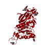
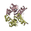
 Rat coronavirus
 X-RAY DIFFRACTION /
  Authors
 Citation
 Structure visualization
 Downloads & links
 Other downloads
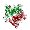

 PDBj
PDBj


 Assembly
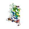
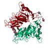
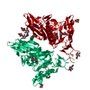
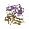
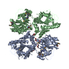
 Homo sapiens (human) / References: UniProt: Q3HS77, sialate O-acetylesterase
 Type: D-saccharide, beta linking / Mass: 221.208 Da / Num. of mol.: 6 / Source method: isolated from a natural source / Formula: C8H15NO6
 Mass: 22.990 Da / Num. of mol.: 1 / Source method: obtained synthetically / Formula: Na
 Mass: 65.409 Da / Num. of mol.: 1 / Source method: obtained synthetically / Formula: Zn
 Mass: 18.015 Da / Num. of mol.: 89 / Source method: isolated from a natural source / Formula: H2O
 Sample preparation
 / Beamline: X06SA / Wavelength: 0.9999 Å
 Processing