 Movie
Movie Controller

[English] 日本語
 Yorodumi
- PDB-4zsb: Crystal structure of the ligand-free effector-binding domain of D... -
+ Open data

- Basic information
| Entry | Database: PDB / ID: 4zsb | ||||||
|---|---|---|---|---|---|---|---|
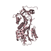
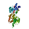
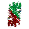
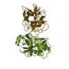
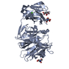
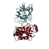
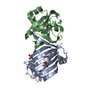
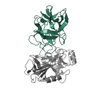
| Title | Crystal structure of the ligand-free effector-binding domain of DasR (DasR-EBD) | ||||||
 Components Components | HTH-type transcriptional repressor DasR | ||||||
 Keywords Keywords | TRANSCRIPTION / Repressor / Bacterial transcription regulation / Transcription factor / GntR/HutC family / Effector-binding domain / N-acetylglucosamine utilization / Ligand-free / Master regulator | ||||||
| Function / homology |  Function and homology information Function and homology informationDNA-binding transcription factor activity / negative regulation of DNA-templated transcription / DNA binding / cytoplasm Similarity search - Function | ||||||
| Biological species | Streptomyces coelicolor A3 | ||||||
| Method |  X-RAY DIFFRACTION / SYNCHROTRON / MOLECULAR REPLACEMENT / Resolution: 2.004 Å X-RAY DIFFRACTION / SYNCHROTRON / MOLECULAR REPLACEMENT / Resolution: 2.004 Å | ||||||
 Authors Authors | Fillenberg, S.B. / Grau, F.C. / Muller, Y.A. | ||||||
| Funding support |  Germany, 1items Germany, 1items
| ||||||
 Citation Citation | Journal: Plos One / Year: 2016 Title: Crystal Structures of the Global Regulator DasR from Streptomyces coelicolor: Implications for the Allosteric Regulation of GntR/HutC Repressors. Authors: Fillenberg, S.B. / Friess, M.D. / Korner, S. / Bockmann, R.A. / Muller, Y.A. | ||||||
| History |
|
- Structure visualization
| Structure viewer | Molecule: MolmilJmol/JSmol |
|---|
- Downloads & links
-Download
| PDBx/mmCIF format | 4zsb.cif.gz | 47.5 KB | Display | PDBx/mmCIF format |
|---|---|---|---|---|
| PDB format | pdb4zsb.ent.gz | 32.2 KB | Display | PDB format |
| PDBx/mmJSON format | 4zsb.json.gz | Tree view | PDBx/mmJSON format | |
| Others |  Other downloads Other downloads |
-Validation report
| Arichive directory | https://data.pdbj.org/pub/pdb/validation_reports/zs/4zsbftp://data.pdbj.org/pub/pdb/validation_reports/zs/4zsb | HTTPS FTP |
|---|
-Related structure data
| Related structure data | 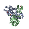 4zs8C 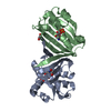 4zsiC  4zskC 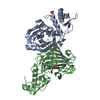 2wv0S S: Starting model for refinement C: citing same article ( |
|---|---|
| Similar structure data |


-Links
 PDBj
PDBj- Assembly
| Deposited unit | 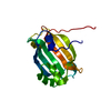
| ||||||||
|---|---|---|---|---|---|---|---|---|---|
| 1 | 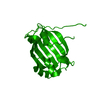
| ||||||||
| Unit cell |
| ||||||||
| Components on special symmetry positions |
|
-Components
| #1: Protein | Mass: 19028.689 Da / Num. of mol.: 1 / Fragment: UNP residues 89-254 Source method: isolated from a genetically manipulated source Details: DasR-EBD comprises residues 88-254 of full-length DasR Source: (gene. exp.)  Streptomyces coelicolor A3(2) (bacteria) Streptomyces coelicolor A3(2) (bacteria)Gene: dasR, SCO5231, SC7E4.28c / Production host: |
|---|---|
| #2: Water | ChemComp-HOH /  Mass: 18.015 Da / Num. of mol.: 75 / Source method: isolated from a natural source / Formula: H2O Mass: 18.015 Da / Num. of mol.: 75 / Source method: isolated from a natural source / Formula: H2O |
-Experimental details
-Experiment
| Experiment | Method: X-RAY DIFFRACTION |
|---|
- Sample preparation
| Crystal | Density Matthews: 2.77 Å3/Da / Density % sol: 55.67 % |
|---|---|
| Crystal grow | Temperature: 292.15 K / Method: vapor diffusion, sitting drop / pH: 9 Details: 0.1 M Bis-Tris propane pH 9.0, 4.0 M potassium formate, 20 % (w/v) PEG monomethyl ether 2,000 |
-Data collection
| Diffraction | Mean temperature: 100 K |
|---|---|
| Diffraction source | Source: SYNCHROTRON / Site: BESSY / Beamline: 14.1 / Wavelength: 0.91841 Å |
| Detector | Type: RAYONIX MX-225 / Detector: CCD / Date: Feb 2, 2011 |
| Radiation | Monochromator: Si-111 crystal / Protocol: SINGLE WAVELENGTH / Monochromatic (M) / Laue (L): M / Scattering type: x-ray |
| Radiation wavelength | Wavelength: 0.91841 Å / Relative weight: 1 |
| Reflection | Resolution: 2→50 Å / Num. obs: 14018 / % possible obs: 98.9 % / Redundancy: 4 % / Rmerge(I) obs: 0.054 / Net I/σ(I): 17.5 |
| Reflection shell | Resolution: 2→2.08 Å / Redundancy: 3 % / Rmerge(I) obs: 0.806 / Mean I/σ(I) obs: 1.4 / % possible all: 94.2 |
- Processing
| Software |
| ||||||||||||||||||||||||||||||||||||||||||||||||||||||||||||||||||||||
|---|---|---|---|---|---|---|---|---|---|---|---|---|---|---|---|---|---|---|---|---|---|---|---|---|---|---|---|---|---|---|---|---|---|---|---|---|---|---|---|---|---|---|---|---|---|---|---|---|---|---|---|---|---|---|---|---|---|---|---|---|---|---|---|---|---|---|---|---|---|---|---|
| Refinement | Method to determine structure: MOLECULAR REPLACEMENT Starting model: Effector-binding domain of entry 2wv0 Resolution: 2.004→44.727 Å / SU ML: 0.3 / Cross valid method: FREE R-VALUE / σ(F): 1.21 / Phase error: 28.46 / Stereochemistry target values: ML
| ||||||||||||||||||||||||||||||||||||||||||||||||||||||||||||||||||||||
| Solvent computation | Shrinkage radii: 0.9 Å / VDW probe radii: 1.11 Å / Solvent model: FLAT BULK SOLVENT MODEL | ||||||||||||||||||||||||||||||||||||||||||||||||||||||||||||||||||||||
| Refinement step | Cycle: LAST / Resolution: 2.004→44.727 Å
| ||||||||||||||||||||||||||||||||||||||||||||||||||||||||||||||||||||||
| Refine LS restraints |
| ||||||||||||||||||||||||||||||||||||||||||||||||||||||||||||||||||||||
| LS refinement shell |
|
