Mass: 18.015 Da / Num. of mol.: 288 / Source method: isolated from a natural source / Formula: H2O
-
Details
Has protein modification
Y
-
Experimental details
-
Experiment
Experiment
Method: X-RAY DIFFRACTION
-
Sample preparation
Crystal
Density Matthews: 2.45 Å3/Da / Density % sol: 51.62 % / Description: Green prismatic crystals.
Crystal grow
Temperature: 298 K / Method: vapor diffusion, hanging drop / pH: 4 Details: Crystals of D207L-Y263F phytochrome mutant were grown from hanging drop vapor diffusion in 20% PEG400, starting protein concentration of 20 mg/ml, and 0.1M phosphate citrate buffer at pH 4.0.
-
Data collection
Diffraction
Mean temperature: 100 K
Diffraction source
Source: ROTATING ANODE / Type: BRUKER AXS MICROSTAR / Wavelength: 1.5418 Å
Detector
Type: BRUKER SMART 6000 / Detector: CCD / Date: Feb 14, 2015
Radiation
Monochromator: Goebel Mirrors / Protocol: SINGLE WAVELENGTH / Monochromatic (M) / Laue (L): M / Scattering type: x-ray
Radiation wavelength
Wavelength: 1.5418 Å / Relative weight: 1
Reflection
Resolution: 1.5→47.6 Å / Num. obs: 54864 / % possible obs: 96 % / Redundancy: 2.9 % / Biso Wilson estimate: 10.84 Å2 / Rsym value: 0.05 / Net I/σ(I): 14.69
Reflection shell
Resolution: 1.5→1.55 Å / Redundancy: 4 % / Rmerge(I) obs: 0.179 / Mean I/σ(I) obs: 4.83 / % possible all: 91.2
-
Processing
Software
Name
Version
Classification
REFMAC
5.8.0107
refinement
SAINT
datareduction
SAINT
datascaling
PHASER
phasing
Refinement
Method to determine structure: MOLECULAR REPLACEMENT Starting model: 4O8G
Resolution: 1.5→25 Å / Cor.coef. Fo:Fc: 0.959 / Cor.coef. Fo:Fc free: 0.943 / SU B: 2.403 / SU ML: 0.041 / Cross valid method: THROUGHOUT / ESU R: 0.082 / ESU R Free: 0.071 / Stereochemistry target values: MAXIMUM LIKELIHOOD / Details: HYDROGENS HAVE BEEN ADDED IN THE RIDING POSITIONS
Rfactor
Num. reflection
% reflection
Selection details
Rfree
0.195
2709
4.9 %
RANDOM
Rwork
0.16
-
-
-
obs
0.162
52043
95.9 %
-
Solvent computation
Ion probe radii: 0.8 Å / Shrinkage radii: 0.8 Å / VDW probe radii: 1.2 Å / Solvent model: BABINET MODEL WITH MASK
 Movie
Movie Controller

 Yorodumi
 Open data
 Basic information
 Components
 Keywords
 Function and homology information
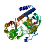
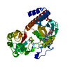
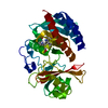
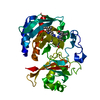
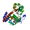
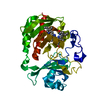
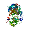
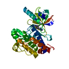
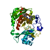
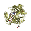
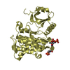
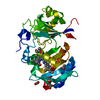
 Deinococcus radiodurans (radioresistant)
 X-RAY DIFFRACTION /
  Authors
 Finland,
  United States, 6items
  Citation
 Structure visualization
 Downloads & links
 Other downloads
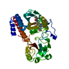
 PDBj
PDBj


 Assembly
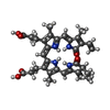
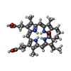


 Mass: 585.670 Da / Num. of mol.: 1 / Source method: obtained synthetically / Formula: C33H37N4O6
 Mass: 585.670 Da / Num. of mol.: 1 / Source method: obtained synthetically / Formula: C33H37N4O6
 Mass: 94.971 Da / Num. of mol.: 1 / Source method: obtained synthetically / Formula: PO4
 Mass: 106.120 Da / Num. of mol.: 2 / Source method: obtained synthetically / Formula: C4H10O3
 Sample preparation
 Processing