| Entry | Database: PDB / ID: 4zqe
|
|---|
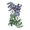
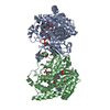
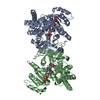
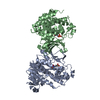
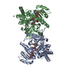
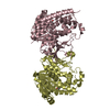
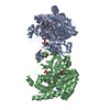
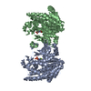
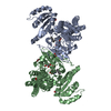
| Title | Crystal structure of DOX-P Reductoisomerase in complex with magnesium |
|---|
 Components Components | 1-deoxy-D-xylulose 5-phosphate reductoisomerase |
|---|
 Keywords Keywords | OXIDOREDUCTASE / MEP pathway / reductoisomerase |
|---|
| Function / homology |  Function and homology information Function and homology information
1-deoxy-D-xylulose-5-phosphate reductoisomerase / 1-deoxy-D-xylulose-5-phosphate reductoisomerase activity / isopentenyl diphosphate biosynthetic process, methylerythritol 4-phosphate pathway / terpenoid biosynthetic process / isomerase activity / NADPH binding / metal ion bindingSimilarity search - Function 1-deoxy-D-xylulose 5-phosphate reductoisomerase / 1-deoxy-D-xylulose 5-phosphate reductoisomerase, N-terminal / 1-deoxy-D-xylulose 5-phosphate reductoisomerase, C-terminal / DXP reductoisomerase C-terminal domain / DXP reductoisomerase, C-terminal domain superfamily / 1-deoxy-D-xylulose 5-phosphate reductoisomerase / 1-deoxy-D-xylulose 5-phosphate reductoisomerase C-terminal domain / DXP reductoisomerase C-terminal domain / RNA polymerase sigma factor, region 2, helix turn helix motif / Rna Polymerase Sigma Factor; Chain: A ...1-deoxy-D-xylulose 5-phosphate reductoisomerase / 1-deoxy-D-xylulose 5-phosphate reductoisomerase, N-terminal / 1-deoxy-D-xylulose 5-phosphate reductoisomerase, C-terminal / DXP reductoisomerase C-terminal domain / DXP reductoisomerase, C-terminal domain superfamily / 1-deoxy-D-xylulose 5-phosphate reductoisomerase / 1-deoxy-D-xylulose 5-phosphate reductoisomerase C-terminal domain / DXP reductoisomerase C-terminal domain / RNA polymerase sigma factor, region 2, helix turn helix motif / Rna Polymerase Sigma Factor; Chain: A / NAD(P)-binding Rossmann-like Domain / NAD(P)-binding domain superfamily / Rossmann fold / Orthogonal Bundle / 3-Layer(aba) Sandwich / Mainly Alpha / Alpha BetaSimilarity search - Domain/homology |
|---|
| Biological species |  Moraxella catarrhalis (bacteria) Moraxella catarrhalis (bacteria) |
|---|
| Method |  X-RAY DIFFRACTION / SYNCHROTRON / MOLECULAR REPLACEMENT / Resolution: 1.98 Å X-RAY DIFFRACTION / SYNCHROTRON / MOLECULAR REPLACEMENT / Resolution: 1.98 Å |
|---|
 Authors Authors | Birkinshaw, R.W. / Brady, R.L. |
|---|
 Citation Citation | Journal: To Be Published
Title: Crystal structures of the Moraxella catarrhalis DOX-P Reductoisomerase
Authors: Birkinshaw, R.W. / Brady, R.L. |
|---|
| History | | Deposition | May 10, 2015 | Deposition site: RCSB / Processing site: PDBE |
|---|
| Revision 1.0 | Jun 29, 2016 | Provider: repository / Type: Initial release |
|---|
| Revision 1.1 | May 8, 2024 | Group: Data collection / Database references / Refinement description
Category: chem_comp_atom / chem_comp_bond ...chem_comp_atom / chem_comp_bond / database_2 / struct_ncs_dom_lim
Item: _database_2.pdbx_DOI / _database_2.pdbx_database_accession ..._database_2.pdbx_DOI / _database_2.pdbx_database_accession / _struct_ncs_dom_lim.beg_auth_comp_id / _struct_ncs_dom_lim.beg_label_asym_id / _struct_ncs_dom_lim.beg_label_comp_id / _struct_ncs_dom_lim.beg_label_seq_id / _struct_ncs_dom_lim.end_auth_comp_id / _struct_ncs_dom_lim.end_label_asym_id / _struct_ncs_dom_lim.end_label_comp_id / _struct_ncs_dom_lim.end_label_seq_id |
|---|
|
|---|
 Movie
Movie Controller

 Yorodumi
 Open data
 Basic information
 Structure visualization
 Downloads & links
 Other downloads
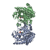


 PDBj
PDBj Assembly
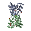

 Mass: 96.063 Da / Num. of mol.: 9 / Source method: obtained synthetically / Formula: SO4
 Mass: 92.094 Da / Num. of mol.: 5 / Source method: obtained synthetically / Formula: C3H8O3
 Mass: 18.015 Da / Num. of mol.: 378 / Source method: isolated from a natural source / Formula: H2O
 Sample preparation
 / Beamline: I04-1 / Wavelength: 0.9173 Å
 Processing