 Movie
Movie Controller

[English] 日本語
 Yorodumi
- PDB-4zj1: Crystal Structure of p-acrylamido-phenylalanine modified TEM1 bet... -
+ Open data

- Basic information
| Entry | Database: PDB / ID: 4zj1 | ||||||
|---|---|---|---|---|---|---|---|
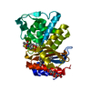
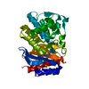
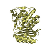
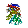
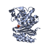
| Title | Crystal Structure of p-acrylamido-phenylalanine modified TEM1 beta-lactamase from Escherichia coli : V216AcrF mutant | ||||||
 Components Components | Beta-lactamase TEM | ||||||
 Keywords Keywords | HYDROLASE / noncanonical amino acid / beta-lactamase / protein evolution / evolutionary advantage | ||||||
| Function / homology |  Function and homology information Function and homology informationAntimicrobial resistance / beta-lactam antibiotic catabolic process / beta-lactamase activity / beta-lactamase / response to antibiotic Similarity search - Function | ||||||
| Biological species |  | ||||||
| Method |  X-RAY DIFFRACTION / MOLECULAR REPLACEMENT / Resolution: 1.54 Å X-RAY DIFFRACTION / MOLECULAR REPLACEMENT / Resolution: 1.54 Å | ||||||
 Authors Authors | Xiao, H. / Nasertorabi, F. / Choi, S. / Han, G.W. / Reed, S.A. / Stevens, R.C. / Schultz, P.G. | ||||||
 Citation Citation | Journal: Proc.Natl.Acad.Sci.USA / Year: 2015 Title: Exploring the potential impact of an expanded genetic code on protein function. Authors: Xiao, H. / Nasertorabi, F. / Choi, S.H. / Han, G.W. / Reed, S.A. / Stevens, R.C. / Schultz, P.G. | ||||||
| History |
|
- Structure visualization
| Structure viewer | Molecule: MolmilJmol/JSmol |
|---|
- Downloads & links
-Download
| PDBx/mmCIF format | 4zj1.cif.gz | 134 KB | Display | PDBx/mmCIF format |
|---|---|---|---|---|
| PDB format | pdb4zj1.ent.gz | 100.6 KB | Display | PDB format |
| PDBx/mmJSON format | 4zj1.json.gz | Tree view | PDBx/mmJSON format | |
| Others |  Other downloads Other downloads |
-Validation report
| Arichive directory | https://data.pdbj.org/pub/pdb/validation_reports/zj/4zj1ftp://data.pdbj.org/pub/pdb/validation_reports/zj/4zj1 | HTTPS FTP |
|---|
-Related structure data
| Related structure data | 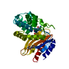 4zj2C 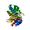 4zj3C  1btlS C: citing same article ( S: Starting model for refinement |
|---|---|
| Similar structure data |


-Links
 PDBj
PDBj


- Assembly
| Deposited unit | 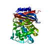
| ||||||||
|---|---|---|---|---|---|---|---|---|---|
| 1 |
| ||||||||
| Unit cell |
|
-Components
| #1: Protein | Mass: 32995.734 Da / Num. of mol.: 1 / Mutation: V216AcrF Source method: isolated from a genetically manipulated source Source: (gene. exp.) |
|---|---|
| #2: Chemical | ChemComp-GOL /   Mass: 92.094 Da / Num. of mol.: 1 / Source method: obtained synthetically / Formula: C3H8O3 Mass: 92.094 Da / Num. of mol.: 1 / Source method: obtained synthetically / Formula: C3H8O3 |
| #3: Chemical | ChemComp-MES /   Mass: 195.237 Da / Num. of mol.: 1 / Source method: obtained synthetically / Formula: C6H13NO4S / Comment: pH buffer*YM Mass: 195.237 Da / Num. of mol.: 1 / Source method: obtained synthetically / Formula: C6H13NO4S / Comment: pH buffer*YM |
| #4: Chemical | ChemComp-PEG /   Mass: 106.120 Da / Num. of mol.: 1 / Source method: obtained synthetically / Formula: C4H10O3 Mass: 106.120 Da / Num. of mol.: 1 / Source method: obtained synthetically / Formula: C4H10O3 |
| #5: Water | ChemComp-HOH /  Mass: 18.015 Da / Num. of mol.: 468 / Source method: isolated from a natural source / Formula: H2O Mass: 18.015 Da / Num. of mol.: 468 / Source method: isolated from a natural source / Formula: H2O |
| Sequence details | The authors used the sequence from NCBI Reference Sequence: WP_000027050.1 |
-Experimental details
-Experiment
| Experiment | Method: X-RAY DIFFRACTION |
|---|
- Sample preparation
| Crystal | Density Matthews: 2.09 Å3/Da / Density % sol: 41.11 % |
|---|---|
| Crystal grow | Temperature: 295 K / Method: vapor diffusion, hanging drop / pH: 7.8 Details: 0.1M 2-(N-morpholino) ethanesulfonic acid (MES) pH 6.5, 15% PEG 20.000 and 15% PEG 550MME |
-Data collection
| Diffraction | Mean temperature: 100 K |
|---|---|
| Diffraction source | Source: ROTATING ANODE / Type: RIGAKU FR-E+ SUPERBRIGHT / Wavelength: 1.5418 Å |
| Detector | Type: RIGAKU RAXIS HTC / Detector: IMAGE PLATE / Date: Sep 4, 2014 |
| Radiation | Protocol: SINGLE WAVELENGTH / Monochromatic (M) / Laue (L): M / Scattering type: x-ray |
| Radiation wavelength | Wavelength: 1.5418 Å / Relative weight: 1 |
| Reflection | Resolution: 1.54→26.54 Å / Num. obs: 41549 / % possible obs: 100 % / Redundancy: 7.4 % / Rmerge(I) obs: 0.064 / Net I/σ(I): 16.3 |
| Reflection shell | Resolution: 1.54→1.63 Å / Redundancy: 7.1 % / Rmerge(I) obs: 0.188 / Mean I/σ(I) obs: 6.5 / % possible all: 100 |
- Processing
| Software |
| ||||||||||||||||||||||||||||||||||||||||||||||||||||||||||||||||||||||||||||||||||||||||||||||||||||||||||||||||||||||||||||||||||||||||||||||||||||||||||||||||||||||||||||||||||||||
|---|---|---|---|---|---|---|---|---|---|---|---|---|---|---|---|---|---|---|---|---|---|---|---|---|---|---|---|---|---|---|---|---|---|---|---|---|---|---|---|---|---|---|---|---|---|---|---|---|---|---|---|---|---|---|---|---|---|---|---|---|---|---|---|---|---|---|---|---|---|---|---|---|---|---|---|---|---|---|---|---|---|---|---|---|---|---|---|---|---|---|---|---|---|---|---|---|---|---|---|---|---|---|---|---|---|---|---|---|---|---|---|---|---|---|---|---|---|---|---|---|---|---|---|---|---|---|---|---|---|---|---|---|---|---|---|---|---|---|---|---|---|---|---|---|---|---|---|---|---|---|---|---|---|---|---|---|---|---|---|---|---|---|---|---|---|---|---|---|---|---|---|---|---|---|---|---|---|---|---|---|---|---|---|
| Refinement | Method to determine structure: MOLECULAR REPLACEMENT Starting model: PDB entry 1BTL Resolution: 1.54→25 Å / Cor.coef. Fo:Fc: 0.974 / Cor.coef. Fo:Fc free: 0.964 / SU B: 2.077 / SU ML: 0.039 / Cross valid method: THROUGHOUT / ESU R: 0.068 / ESU R Free: 0.07 / Stereochemistry target values: MAXIMUM LIKELIHOOD / Details: HYDROGENS HAVE BEEN ADDED IN THE RIDING POSITIONS
| ||||||||||||||||||||||||||||||||||||||||||||||||||||||||||||||||||||||||||||||||||||||||||||||||||||||||||||||||||||||||||||||||||||||||||||||||||||||||||||||||||||||||||||||||||||||
| Solvent computation | Ion probe radii: 0.8 Å / Shrinkage radii: 0.8 Å / VDW probe radii: 1.2 Å / Solvent model: MASK | ||||||||||||||||||||||||||||||||||||||||||||||||||||||||||||||||||||||||||||||||||||||||||||||||||||||||||||||||||||||||||||||||||||||||||||||||||||||||||||||||||||||||||||||||||||||
| Displacement parameters | Biso mean: 16.414 Å2
| ||||||||||||||||||||||||||||||||||||||||||||||||||||||||||||||||||||||||||||||||||||||||||||||||||||||||||||||||||||||||||||||||||||||||||||||||||||||||||||||||||||||||||||||||||||||
| Refinement step | Cycle: 1 / Resolution: 1.54→25 Å
| ||||||||||||||||||||||||||||||||||||||||||||||||||||||||||||||||||||||||||||||||||||||||||||||||||||||||||||||||||||||||||||||||||||||||||||||||||||||||||||||||||||||||||||||||||||||
| Refine LS restraints |
|