Mass: 18.015 Da / Num. of mol.: 102 / Source method: isolated from a natural source / Formula: H2O
Has protein modification
Y
Sequence details
THE CONSTRUCT (4-86) WAS EXPRESSED WITH A PURIFICATION TAG MGSDKIHHHHHHENLYFQG. THE TAG WAS REMOVED ...THE CONSTRUCT (4-86) WAS EXPRESSED WITH A PURIFICATION TAG MGSDKIHHHHHHENLYFQG. THE TAG WAS REMOVED WITH TEV PROTEASE LEAVING ONLY A GLYCINE (0) FOLLOWED BY THE TARGET SEQUENCE.
-
Experimental details
-
Experiment
Experiment
Method: X-RAY DIFFRACTION / Number of used crystals: 1
-
Sample preparation
Crystal
Density Matthews: 2.08 Å3/Da / Density % sol: 40.83 %
Crystal grow
Temperature: 293 K / Method: vapor diffusion, sitting drop / Details: 25.0% Glycerol, 1.50M ammonium sulfate
Resolution: 1.5→27.366 Å / Num. obs: 12873 / % possible obs: 92.4 % / Observed criterion σ(I): -3 / Redundancy: 1.685 % / Biso Wilson estimate: 17.415 Å2 / Rmerge F obs: 0.984 / Rmerge(I) obs: 0.092 / Rrim(I) all: 0.128 / Net I/σ(I): 7.56 / Num. measured all: 39652
Reflection shell
Resolution (Å)
Highest resolution (Å)
Rmerge F obs
Rmerge(I) obs
Mean I/σ(I) obs
Num. measured obs
Num. possible
Num. unique obs
Rrim(I) all
Diffraction-ID
% possible all
1.5-1.55
0.782
0.392
1.8
3742
2425
2234
0.538
1
92.1
1.55-1.62
0.86
0.32
2
4494
2851
2661
0.439
93.3
1.62-1.69
0.907
0.252
2.6
3697
2353
2188
0.346
93
1.69-1.78
0.92
0.215
3.2
4090
2605
2432
0.297
93.4
1.78-1.89
0.962
0.153
4.3
3888
2510
2319
0.211
92.4
1.89-2.04
0.966
0.122
5.8
3937
2586
2363
0.171
91.4
2.04-2.24
0.979
0.086
8.6
3605
2467
2193
0.121
88.9
2.24-2.56
0.958
0.102
11.4
3869
2542
2305
0.143
90.7
2.56-3.23
0.942
0.111
15.3
4294
2580
2491
0.155
96.6
3.23
0.986
0.057
20.4
4036
2555
2341
0.08
91.6
-
Phasing
Phasing
Method: MAD
-
Processing
Software
Name
Version
Classification
PDB_EXTRACT
3.1
dataextraction
SOLVE
phasing
XSCALE
November3, 2014BUILT=20141118
datascaling
BUSTER-TNT
2.10.2
refinement
BUSTER
2.10.2
refinement
XDS
datareduction
Refinement
Method to determine structure: MAD / Resolution: 1.5→27.366 Å / Cor.coef. Fo:Fc: 0.9483 / Cor.coef. Fo:Fc free: 0.9332 / Occupancy max: 1 / Occupancy min: 0.5 / Cross valid method: THROUGHOUT / σ(F): 0 Details: 1. ATOM RECORDS CONTAIN SUM OF TLS AND RESIDUAL B FACTORS. 2. ANISOU RECORDS CONTAIN SUM OF TLS AND RESIDUAL U FACTORS. 3. A MET-INHIBITION PROTOCOL WAS USED FOR SELENOMETHIONINE ...Details: 1. ATOM RECORDS CONTAIN SUM OF TLS AND RESIDUAL B FACTORS. 2. ANISOU RECORDS CONTAIN SUM OF TLS AND RESIDUAL U FACTORS. 3. A MET-INHIBITION PROTOCOL WAS USED FOR SELENOMETHIONINE INCORPORATION DURING PROTEIN EXPRESSION. THE OCCUPANCY OF THE SE ATOMS IN THE MSE RESIDUES WAS REDUCED TO 0.75 FOR THE REDUCED SCATTERING POWER DUE TO PARTIAL S-MET INCORPORATION. 4. THE MAD PHASES WERE USED AS RESTRAINTS DURING REFINEMENT
In the structure databanks used in Yorodumi, some data are registered as the other names, "COVID-19 virus" and "2019-nCoV". Here are the details of the virus and the list of structure data.
Jan 31, 2019. EMDB accession codes are about to change! (news from PDBe EMDB page)
EMDB accession codes are about to change! (news from PDBe EMDB page)
The allocation of 4 digits for EMDB accession codes will soon come to an end. Whilst these codes will remain in use, new EMDB accession codes will include an additional digit and will expand incrementally as the available range of codes is exhausted. The current 4-digit format prefixed with “EMD-” (i.e. EMD-XXXX) will advance to a 5-digit format (i.e. EMD-XXXXX), and so on. It is currently estimated that the 4-digit codes will be depleted around Spring 2019, at which point the 5-digit format will come into force.
The EM Navigator/Yorodumi systems omit the EMD- prefix.
Related info.:Q: What is EMD? / ID/Accession-code notation in Yorodumi/EM Navigator
Yorodumi is a browser for structure data from EMDB, PDB, SASBDB, etc.
This page is also the successor to EM Navigator detail page, and also detail information page/front-end page for Omokage search.
The word "yorodu" (or yorozu) is an old Japanese word meaning "ten thousand". "mi" (miru) is to see.
Related info.:EMDB / PDB / SASBDB / Comparison of 3 databanks / Yorodumi Search / Aug 31, 2016. New EM Navigator & Yorodumi / Yorodumi Papers / Jmol/JSmol / Function and homology information / Changes in new EM Navigator and Yorodumi
 Movie
Movie Controller

 Yorodumi
 Open data
 Basic information
 Components
 Keywords
 Function and homology information
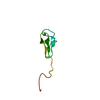
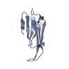
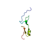
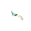
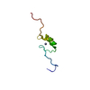
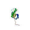
 Homo sapiens (human)
 X-RAY DIFFRACTION /
  Authors
 Citation
 Structure visualization
 Downloads & links
 Other downloads
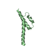

 PDBj
PDBj Assembly
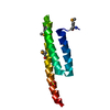


 Mass: 96.063 Da / Num. of mol.: 2 / Source method: obtained synthetically / Formula: SO4
 Mass: 18.015 Da / Num. of mol.: 102 / Source method: isolated from a natural source / Formula: H2O
 Sample preparation
 / Beamline: BL14-1 / Wavelength: 0.95369,0.97968,0.97943
 Processing