 Movie
Movie Controller

[English] 日本語
 Yorodumi
- PDB-4ze5: Structure of Gan1D-E170Q, a catalytic mutant of a putative 6-phos... -
+ Open data

- Basic information
| Entry | Database: PDB / ID: 4ze5 | ||||||
|---|---|---|---|---|---|---|---|
| Title | Structure of Gan1D-E170Q, a catalytic mutant of a putative 6-phospho-beta-galactosidase from Geobacillus stearothermophilus | ||||||
 Components Components | Putative 6-phospho-beta-galactobiosidase | ||||||
 Keywords Keywords | HYDROLASE / TIM-barrel / catalytic mutant / glycoside hydrolase / 6-phospho-beta-galactosidase | ||||||
| Function / homology |  Function and homology information Function and homology information6-phospho-beta-galactosidase / 6-phospho-beta-galactosidase activity / carbohydrate catabolic process / beta-glucosidase activity / cytosol Similarity search - Function | ||||||
| Biological species |   Geobacillus stearothermophilus (bacteria) Geobacillus stearothermophilus (bacteria) | ||||||
| Method |  X-RAY DIFFRACTION / SYNCHROTRON / MOLECULAR REPLACEMENT / Resolution: 1.48 Å X-RAY DIFFRACTION / SYNCHROTRON / MOLECULAR REPLACEMENT / Resolution: 1.48 Å | ||||||
 Authors Authors | Lansky, S. / Zehavi, A. / Dvir, H. / Shoham, Y. / Shoham, G. | ||||||
 Citation Citation | Journal: To Be Published Title: Structure of Gan1D-E170Q, a catalytic mutant of a putative 6-phospho-beta-galactosidase from Geobacillus stearothermophilus Authors: Lansky, S. / Zehavi, A. / Dvir, H. / Shoham, Y. / Shoham, G. | ||||||
| History |
|
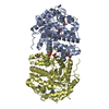
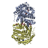
- Structure visualization
| Structure viewer | Molecule: MolmilJmol/JSmol |
|---|
- Downloads & links
-Download
| PDBx/mmCIF format | 4ze5.cif.gz | 453.5 KB | Display | PDBx/mmCIF format |
|---|---|---|---|---|
| PDB format | pdb4ze5.ent.gz | 364.2 KB | Display | PDB format |
| PDBx/mmJSON format | 4ze5.json.gz | Tree view | PDBx/mmJSON format | |
| Others |  Other downloads Other downloads |
-Validation report
| Arichive directory | https://data.pdbj.org/pub/pdb/validation_reports/ze/4ze5ftp://data.pdbj.org/pub/pdb/validation_reports/ze/4ze5 | HTTPS FTP |
|---|
-Related structure data
| Related structure data |  4zeh S: Starting model for refinement |
|---|---|
| Similar structure data |


-Links
 PDBj
PDBj


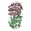
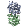
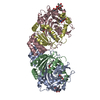
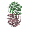
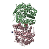
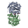
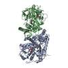
- Assembly
| Deposited unit | 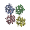
| ||||||||
|---|---|---|---|---|---|---|---|---|---|
| 1 | 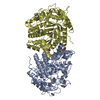
| ||||||||
| 2 | 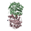
| ||||||||
| 3 | 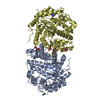
| ||||||||
| 4 | 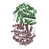
| ||||||||
| Unit cell |
|
-Components
| #1: Protein | Mass: 56208.730 Da / Num. of mol.: 4 Source method: isolated from a genetically manipulated source Source: (gene. exp.) Geobacillus stearothermophilus (bacteria)Gene: gan1D / Production host: #2: Chemical | ChemComp-GOL /   Mass: 92.094 Da / Num. of mol.: 10 / Source method: obtained synthetically / Formula: C3H8O3 Mass: 92.094 Da / Num. of mol.: 10 / Source method: obtained synthetically / Formula: C3H8O3#3: Chemical |   Mass: 69.085 Da / Num. of mol.: 3 / Source method: isolated from a natural source / Formula: C3H5N2 Mass: 69.085 Da / Num. of mol.: 3 / Source method: isolated from a natural source / Formula: C3H5N2#4: Water | ChemComp-HOH / |  Mass: 18.015 Da / Num. of mol.: 2556 / Source method: isolated from a natural source / Formula: H2O Mass: 18.015 Da / Num. of mol.: 2556 / Source method: isolated from a natural source / Formula: H2O |
|---|
-Experimental details
-Experiment
| Experiment | Method: X-RAY DIFFRACTION |
|---|
- Sample preparation
| Crystal | Density Matthews: 2.29 Å3/Da / Density % sol: 46.39 % |
|---|---|
| Crystal grow | Temperature: 293 K / Method: vapor diffusion, hanging drop / pH: 6.5 Details: 16-18% PEG 8K, 3% MPD, 0.1 M imidazole buffer pH 6.5 |
-Data collection
| Diffraction | Mean temperature: 100 K |
|---|---|
| Diffraction source | Source: SYNCHROTRON / Site: ESRF  / Beamline: BM14 / Wavelength: 0.98 Å / Beamline: BM14 / Wavelength: 0.98 Å |
| Detector | Type: MARMOSAIC 225 mm CCD / Detector: CCD / Date: Apr 10, 2014 |
| Radiation | Protocol: SINGLE WAVELENGTH / Monochromatic (M) / Laue (L): M / Scattering type: x-ray |
| Radiation wavelength | Wavelength: 0.98 Å / Relative weight: 1 |
| Reflection | Resolution: 1.48→35 Å / Num. obs: 337040 / % possible obs: 99.7 % / Redundancy: 5.7 % / Rmerge(I) obs: 0.067 / Net I/σ(I): 12.6 |
| Reflection shell | Resolution: 1.48→1.51 Å / Redundancy: 5 % / Rmerge(I) obs: 0.375 / Mean I/σ(I) obs: 3.9 / % possible all: 94.9 |
- Processing
| Software |
| ||||||||||||||||||||||||||||||||||||||||||||||||||||||||||||||||||||||||||||||||||||||||||||||||||||||||||||||||||||||||||||||||||||||||||||||||||||||||||||||||||||||||||||||||||||||
|---|---|---|---|---|---|---|---|---|---|---|---|---|---|---|---|---|---|---|---|---|---|---|---|---|---|---|---|---|---|---|---|---|---|---|---|---|---|---|---|---|---|---|---|---|---|---|---|---|---|---|---|---|---|---|---|---|---|---|---|---|---|---|---|---|---|---|---|---|---|---|---|---|---|---|---|---|---|---|---|---|---|---|---|---|---|---|---|---|---|---|---|---|---|---|---|---|---|---|---|---|---|---|---|---|---|---|---|---|---|---|---|---|---|---|---|---|---|---|---|---|---|---|---|---|---|---|---|---|---|---|---|---|---|---|---|---|---|---|---|---|---|---|---|---|---|---|---|---|---|---|---|---|---|---|---|---|---|---|---|---|---|---|---|---|---|---|---|---|---|---|---|---|---|---|---|---|---|---|---|---|---|---|---|
| Refinement | Method to determine structure: MOLECULAR REPLACEMENT Starting model: 4ZEH 4zeh Resolution: 1.48→27.64 Å / Cor.coef. Fo:Fc: 0.979 / Cor.coef. Fo:Fc free: 0.97 / SU B: 0.858 / SU ML: 0.033 / Cross valid method: THROUGHOUT / ESU R: 0.054 / ESU R Free: 0.058 / Stereochemistry target values: MAXIMUM LIKELIHOOD / Details: HYDROGENS HAVE BEEN ADDED IN THE RIDING POSITIONS
| ||||||||||||||||||||||||||||||||||||||||||||||||||||||||||||||||||||||||||||||||||||||||||||||||||||||||||||||||||||||||||||||||||||||||||||||||||||||||||||||||||||||||||||||||||||||
| Solvent computation | Ion probe radii: 0.8 Å / Shrinkage radii: 0.8 Å / VDW probe radii: 1.2 Å / Solvent model: MASK | ||||||||||||||||||||||||||||||||||||||||||||||||||||||||||||||||||||||||||||||||||||||||||||||||||||||||||||||||||||||||||||||||||||||||||||||||||||||||||||||||||||||||||||||||||||||
| Displacement parameters | Biso mean: 14.232 Å2
| ||||||||||||||||||||||||||||||||||||||||||||||||||||||||||||||||||||||||||||||||||||||||||||||||||||||||||||||||||||||||||||||||||||||||||||||||||||||||||||||||||||||||||||||||||||||
| Refinement step | Cycle: LAST / Resolution: 1.48→27.64 Å
| ||||||||||||||||||||||||||||||||||||||||||||||||||||||||||||||||||||||||||||||||||||||||||||||||||||||||||||||||||||||||||||||||||||||||||||||||||||||||||||||||||||||||||||||||||||||
| Refine LS restraints |
|