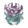
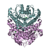
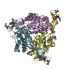
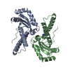
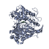
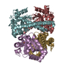
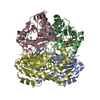
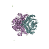
A: Halohydrin epoxidase B B: Halohydrin epoxidase B C: Halohydrin epoxidase B D: Halohydrin epoxidase B E: Halohydrin epoxidase B F: Halohydrin epoxidase B hetero molecules
Protocol: SINGLE WAVELENGTH / Monochromatic (M) / Laue (L): M / Scattering type: x-ray
Radiation wavelength
Wavelength: 1 Å / Relative weight: 1
Reflection
Resolution: 1.6→50 Å / Num. obs: 158218 / % possible obs: 87.8 % / Redundancy: 4.1 % / Rmerge(I) obs: 0.058 / Net I/σ(I): 21.7
Reflection shell
Resolution: 1.6→1.66 Å / Redundancy: 3.9 % / Rmerge(I) obs: 0.271 / Mean I/σ(I) obs: 3.86 / % possible all: 97.7
-
Processing
Software
Name
Version
Classification
PHENIX
(1.10-2152-000)
refinement
HKL-2000
datareduction
HKL-2000
datascaling
SOLVE
phasing
Refinement
Method to determine structure: SAD / Resolution: 1.6→35.31 Å / SU ML: 0.15 / Cross valid method: THROUGHOUT / σ(F): 1.34 / Phase error: 18.96 / Stereochemistry target values: ML
Rfactor
Num. reflection
% reflection
Selection details
Rfree
0.2065
7926
5.02 %
RANDOM
Rwork
0.175
-
-
-
obs
0.1766
157936
87.57 %
-
Solvent computation
Shrinkage radii: 0.9 Å / VDW probe radii: 1.11 Å / Solvent model: FLAT BULK SOLVENT MODEL
Refinement step
Cycle: LAST / Resolution: 1.6→35.31 Å
Protein
Nucleic acid
Ligand
Solvent
Total
Num. atoms
10596
0
21
1045
11662
Refine LS restraints
Refine-ID
Type
Dev ideal
Number
X-RAY DIFFRACTION
f_bond_d
0.006
10853
X-RAY DIFFRACTION
f_angle_d
0.791
14746
X-RAY DIFFRACTION
f_dihedral_angle_d
14.758
6353
X-RAY DIFFRACTION
f_chiral_restr
0.053
1620
X-RAY DIFFRACTION
f_plane_restr
0.006
1943
LS refinement shell
Resolution (Å)
Rfactor Rfree
Num. reflection Rfree
Rfactor Rwork
Num. reflection Rwork
Refine-ID
% reflection obs (%)
1.5999-1.6181
0.2365
302
0.197
5260
X-RAY DIFFRACTION
93
1.6181-1.6372
0.2533
292
0.1879
5603
X-RAY DIFFRACTION
99
1.6372-1.6571
0.2315
340
0.1898
5580
X-RAY DIFFRACTION
99
1.6571-1.6781
0.2083
300
0.1797
5626
X-RAY DIFFRACTION
100
1.6781-1.7002
0.2253
321
0.1742
5628
X-RAY DIFFRACTION
100
1.7002-1.7235
0.2157
301
0.167
5687
X-RAY DIFFRACTION
100
1.7235-1.7481
0.2195
270
0.1669
5664
X-RAY DIFFRACTION
100
1.7481-1.7742
0.2265
288
0.166
5657
X-RAY DIFFRACTION
100
1.7742-1.8019
0.2189
313
0.1675
5673
X-RAY DIFFRACTION
100
1.8019-1.8314
0.2163
292
0.1685
5647
X-RAY DIFFRACTION
100
1.8314-1.863
0.2347
276
0.1837
5697
X-RAY DIFFRACTION
100
1.863-1.8969
0.3237
155
0.2558
2911
X-RAY DIFFRACTION
91
1.8969-1.9334
0.5996
38
0.5174
1078
X-RAY DIFFRACTION
60
1.9334-1.9728
0.3381
250
0.2743
4772
X-RAY DIFFRACTION
92
1.9728-2.0157
0.2158
290
0.1821
5659
X-RAY DIFFRACTION
100
2.0157-2.0626
0.1979
289
0.1679
5696
X-RAY DIFFRACTION
100
2.0626-2.1142
0.2155
281
0.1652
5711
X-RAY DIFFRACTION
100
2.1142-2.1714
0.1954
311
0.1681
5699
X-RAY DIFFRACTION
100
2.1714-2.2352
0.2211
153
0.1955
2664
X-RAY DIFFRACTION
88
2.2352-2.3074
0.2281
65
0.2072
1112
X-RAY DIFFRACTION
80
2.3074-2.3898
0.1901
282
0.1718
5730
X-RAY DIFFRACTION
100
2.3898-2.4855
0.2055
324
0.1666
5662
X-RAY DIFFRACTION
100
2.4855-2.5986
0.1893
336
0.1721
5650
X-RAY DIFFRACTION
100
2.5986-2.7355
0.2021
306
0.1722
5697
X-RAY DIFFRACTION
100
2.7355-2.9068
0.2335
291
0.1744
5728
X-RAY DIFFRACTION
99
2.9068-3.1312
0.1911
286
0.1748
5717
X-RAY DIFFRACTION
99
3.1312-3.446
0.2153
293
0.1724
5728
X-RAY DIFFRACTION
99
3.446-3.9441
0.1732
120
0.1659
2238
X-RAY DIFFRACTION
44
3.9441-4.9669
0.1527
258
0.1382
5251
X-RAY DIFFRACTION
96
4.9669-35.3188
0.1747
303
0.157
5585
X-RAY DIFFRACTION
93
+
About Yorodumi
-
News
-
Feb 9, 2022. New format data for meta-information of EMDB entries
New format data for meta-information of EMDB entries
Version 3 of the EMDB header file is now the official format.
The previous official version 1.9 will be removed from the archive.
In the structure databanks used in Yorodumi, some data are registered as the other names, "COVID-19 virus" and "2019-nCoV". Here are the details of the virus and the list of structure data.
Jan 31, 2019. EMDB accession codes are about to change! (news from PDBe EMDB page)
EMDB accession codes are about to change! (news from PDBe EMDB page)
The allocation of 4 digits for EMDB accession codes will soon come to an end. Whilst these codes will remain in use, new EMDB accession codes will include an additional digit and will expand incrementally as the available range of codes is exhausted. The current 4-digit format prefixed with “EMD-” (i.e. EMD-XXXX) will advance to a 5-digit format (i.e. EMD-XXXXX), and so on. It is currently estimated that the 4-digit codes will be depleted around Spring 2019, at which point the 5-digit format will come into force.
The EM Navigator/Yorodumi systems omit the EMD- prefix.
Related info.:Q: What is EMD? / ID/Accession-code notation in Yorodumi/EM Navigator
Yorodumi is a browser for structure data from EMDB, PDB, SASBDB, etc.
This page is also the successor to EM Navigator detail page, and also detail information page/front-end page for Omokage search.
The word "yorodu" (or yorozu) is an old Japanese word meaning "ten thousand". "mi" (miru) is to see.
Related info.:EMDB / PDB / SASBDB / Comparison of 3 databanks / Yorodumi Search / Aug 31, 2016. New EM Navigator & Yorodumi / Yorodumi Papers / Jmol/JSmol / Function and homology information / Changes in new EM Navigator and Yorodumi
 Movie
Movie Controller


 Open data
 Basic information
 Components
 Keywords
 Function and homology information
 Corynebacterium sp. (bacteria)
 X-RAY DIFFRACTION /
  Authors
 Citation
 Structure visualization
 Downloads & links
 Other downloads


 PDBj
PDBj

 Assembly
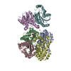

 Mass: 35.453 Da / Num. of mol.: 6 / Source method: obtained synthetically / Formula: Cl
 Mass: 195.237 Da / Num. of mol.: 1 / Source method: obtained synthetically / Formula: C6H13NO4S / Comment: pH buffer*YM
 Mass: 24.305 Da / Num. of mol.: 3 / Source method: obtained synthetically / Formula: Mg
 Mass: 18.015 Da / Num. of mol.: 1045 / Source method: isolated from a natural source / Formula: H2O
 Sample preparation
 / Beamline: BL-5A / Wavelength: 1 Å
 Processing