 Movie
Movie Controller


+ Open data

- Basic information
| Entry | Database: PDB / ID: 4zcb | ||||||
|---|---|---|---|---|---|---|---|
| Title | Human CRBPII mutant - Y60W dimer | ||||||
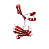
 Components Components | Retinol-binding protein 2 | ||||||
 Keywords Keywords | LIPID BINDING PROTEIN / RETINOL-BINDING PROTEIN / DOMAIN SWAPPING DIMERIZATION | ||||||
| Function / homology |  Function and homology information Function and homology informationvitamin A metabolic process / triglyceride biosynthetic process / retinoid binding / retinal binding / molecular carrier activity / retinol binding / epidermis development / fatty acid transport / Retinoid metabolism and transport / fatty acid binding ...vitamin A metabolic process / triglyceride biosynthetic process / retinoid binding / retinal binding / molecular carrier activity / retinol binding / epidermis development / fatty acid transport / Retinoid metabolism and transport / fatty acid binding / nucleus / cytosol Similarity search - Function | ||||||
| Biological species |  Homo sapiens (human) Homo sapiens (human) | ||||||
| Method |  X-RAY DIFFRACTION / SYNCHROTRON / MOLECULAR REPLACEMENT / Resolution: 1.7 Å X-RAY DIFFRACTION / SYNCHROTRON / MOLECULAR REPLACEMENT / Resolution: 1.7 Å | ||||||
 Authors Authors | Nossoni, Z. / Assar, Z. / Wang, W. / Geiger, J. / Borhan, B. | ||||||
 Citation Citation | Journal: Structure / Year: 2016 Title: Domain-Swapped Dimers of Intracellular Lipid-Binding Proteins: Evidence for Ordered Folding Intermediates. Authors: Assar, Z. / Nossoni, Z. / Wang, W. / Santos, E.M. / Kramer, K. / McCornack, C. / Vasileiou, C. / Borhan, B. / Geiger, J.H. #1: Journal: Acta Crystallogr.,Sect.D / Year: 2014Title: Structures of holo wild-type human cellular retinol-binding protein II (hCRBPII) bound to retinol and retinal. Authors: Nossoni, Z. / Assar, Z. / Yapici, I. / Nosrati, M. / Wang, W. / Berbasova, T. / Vasileiou, C. / Borhan, B. / Geiger, J. | ||||||
| History |
|
- Structure visualization
| Structure viewer | Molecule: MolmilJmol/JSmol |
|---|
- Downloads & links
-Download
| PDBx/mmCIF format | 4zcb.cif.gz | 73.4 KB | Display | PDBx/mmCIF format |
|---|---|---|---|---|
| PDB format | pdb4zcb.ent.gz | 53.5 KB | Display | PDB format |
| PDBx/mmJSON format | 4zcb.json.gz | Tree view | PDBx/mmJSON format | |
| Others |  Other downloads Other downloads |
-Validation report
| Arichive directory | https://data.pdbj.org/pub/pdb/validation_reports/zc/4zcbftp://data.pdbj.org/pub/pdb/validation_reports/zc/4zcb | HTTPS FTP |
|---|
-Related structure data
| Related structure data |  4zguC  4zh6C  4zh9C 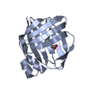 4zj0C  4zr2C  5dg4C  5dpqC  4qynS S: Starting model for refinement C: citing same article ( |
|---|---|
| Similar structure data |


-Links
 PDBj
PDBj


- Assembly
| Deposited unit | 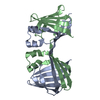
| ||||||||
|---|---|---|---|---|---|---|---|---|---|
| 1 |
| ||||||||
| Unit cell |
|
-Components
| #1: Protein | Mass: 15620.487 Da / Num. of mol.: 2 / Fragment: UNP residues 2-134 / Mutation: Y60W Source method: isolated from a genetically manipulated source Source: (gene. exp.) Homo sapiens (human) / Gene: RBP2, CRBP2 / Plasmid: pET17b / Production host:  #2: Water | ChemComp-HOH / |  Mass: 18.015 Da / Num. of mol.: 217 / Source method: isolated from a natural source / Formula: H2O Mass: 18.015 Da / Num. of mol.: 217 / Source method: isolated from a natural source / Formula: H2O |
|---|
-Experimental details
-Experiment
| Experiment | Method: X-RAY DIFFRACTION |
|---|
- Sample preparation
| Crystal | Density Matthews: 2 Å3/Da / Density % sol: 38.44 % |
|---|---|
| Crystal grow | Temperature: 294 K / Method: vapor diffusion, hanging drop / pH: 4.6 Details: 30% PEG 4000, 0.1M CH3COONa.3H2O, pH 4.6, 0.1M CH3COONH4 |
-Data collection
| Diffraction | Mean temperature: 200 K |
|---|---|
| Diffraction source | Source: SYNCHROTRON / Site: APS  / Beamline: 21-ID-D / Wavelength: 1.1271 Å / Beamline: 21-ID-D / Wavelength: 1.1271 Å |
| Detector | Type: MARMOSAIC 300 mm CCD / Detector: CCD / Date: Oct 22, 2012 |
| Radiation | Protocol: SINGLE WAVELENGTH / Monochromatic (M) / Laue (L): M / Scattering type: x-ray |
| Radiation wavelength | Wavelength: 1.1271 Å / Relative weight: 1 |
| Reflection | Resolution: 1.7→50 Å / Num. obs: 26934 / % possible obs: 98.6 % / Redundancy: 5.2 % / Net I/σ(I): 66.13 |
| Reflection shell | Highest resolution: 1.7 Å / Mean I/σ(I) obs: 7.71 |
- Processing
| Software |
| ||||||||||||||||||||||||||||||||||||||||||||||||||||||||||||||||||||||||||||||||||||||||||||||||||||||||||||||||||||||||||||||||||||||||||||||||||||||||||||||||||||||||||||||||||||||
|---|---|---|---|---|---|---|---|---|---|---|---|---|---|---|---|---|---|---|---|---|---|---|---|---|---|---|---|---|---|---|---|---|---|---|---|---|---|---|---|---|---|---|---|---|---|---|---|---|---|---|---|---|---|---|---|---|---|---|---|---|---|---|---|---|---|---|---|---|---|---|---|---|---|---|---|---|---|---|---|---|---|---|---|---|---|---|---|---|---|---|---|---|---|---|---|---|---|---|---|---|---|---|---|---|---|---|---|---|---|---|---|---|---|---|---|---|---|---|---|---|---|---|---|---|---|---|---|---|---|---|---|---|---|---|---|---|---|---|---|---|---|---|---|---|---|---|---|---|---|---|---|---|---|---|---|---|---|---|---|---|---|---|---|---|---|---|---|---|---|---|---|---|---|---|---|---|---|---|---|---|---|---|---|
| Refinement | Method to determine structure: MOLECULAR REPLACEMENT Starting model: PDB entry 4QYN Resolution: 1.7→31.59 Å / Cor.coef. Fo:Fc: 0.959 / Cor.coef. Fo:Fc free: 0.937 / SU B: 2.051 / SU ML: 0.071 / Cross valid method: THROUGHOUT / ESU R: 0.118 / ESU R Free: 0.124 / Stereochemistry target values: MAXIMUM LIKELIHOOD / Details: HYDROGENS HAVE BEEN ADDED IN THE RIDING POSITIONS
| ||||||||||||||||||||||||||||||||||||||||||||||||||||||||||||||||||||||||||||||||||||||||||||||||||||||||||||||||||||||||||||||||||||||||||||||||||||||||||||||||||||||||||||||||||||||
| Solvent computation | Ion probe radii: 0.8 Å / Shrinkage radii: 0.8 Å / VDW probe radii: 1.4 Å / Solvent model: MASK | ||||||||||||||||||||||||||||||||||||||||||||||||||||||||||||||||||||||||||||||||||||||||||||||||||||||||||||||||||||||||||||||||||||||||||||||||||||||||||||||||||||||||||||||||||||||
| Displacement parameters | Biso mean: 22.498 Å2
| ||||||||||||||||||||||||||||||||||||||||||||||||||||||||||||||||||||||||||||||||||||||||||||||||||||||||||||||||||||||||||||||||||||||||||||||||||||||||||||||||||||||||||||||||||||||
| Refinement step | Cycle: 1 / Resolution: 1.7→31.59 Å
| ||||||||||||||||||||||||||||||||||||||||||||||||||||||||||||||||||||||||||||||||||||||||||||||||||||||||||||||||||||||||||||||||||||||||||||||||||||||||||||||||||||||||||||||||||||||
| Refine LS restraints |
|