 Movie
Movie Controller


+ Open data

- Basic information
| Entry | Database: PDB / ID: 4yxt | ||||||
|---|---|---|---|---|---|---|---|
| Title | PksG, a HMG-CoA Synthase from Bacillus subtilus | ||||||
 Components Components | Polyketide biosynthesis 3-hydroxy-3-methylglutaryl-ACP synthase PksG | ||||||
 Keywords Keywords | TRANSFERASE / bacillaene / polyketide | ||||||
| Function / homology |  Function and homology information Function and homology informationTransferases; Acyltransferases; Acyl groups converted into alkyl groups on transfer / hydroxymethylglutaryl-CoA synthase activity / farnesyl diphosphate biosynthetic process, mevalonate pathway / acetyl-CoA metabolic process / antibiotic biosynthetic process / cytoplasm Similarity search - Function | ||||||
| Biological species |  | ||||||
| Method |  X-RAY DIFFRACTION / SYNCHROTRON / MOLECULAR REPLACEMENT / molecular replacement / Resolution: 2.1 Å X-RAY DIFFRACTION / SYNCHROTRON / MOLECULAR REPLACEMENT / molecular replacement / Resolution: 2.1 Å | ||||||
 Authors Authors | Till, M. / Nair, A.V. / Robson, A. / Race, P.R. | ||||||
 Citation Citation | Journal: To Be Published Title: PksG, a HMG-CoA Synthase from Bacillus subtilus Authors: Till, M. / Nair, A.V. / Robson, A. / Race, P.R. | ||||||
| History |
|
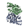
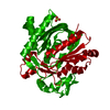
- Structure visualization
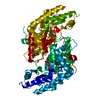
| Structure viewer | Molecule: MolmilJmol/JSmol |
|---|
- Downloads & links
-Download
| PDBx/mmCIF format | 4yxt.cif.gz | 167.4 KB | Display | PDBx/mmCIF format |
|---|---|---|---|---|
| PDB format | pdb4yxt.ent.gz | 132.4 KB | Display | PDB format |
| PDBx/mmJSON format | 4yxt.json.gz | Tree view | PDBx/mmJSON format | |
| Others |  Other downloads Other downloads |
-Validation report
| Arichive directory | https://data.pdbj.org/pub/pdb/validation_reports/yx/4yxtftp://data.pdbj.org/pub/pdb/validation_reports/yx/4yxt | HTTPS FTP |
|---|
-Related structure data


-Links
 PDBj
PDBj

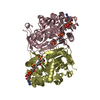
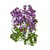
- Assembly
| Deposited unit | 
| ||||||||
|---|---|---|---|---|---|---|---|---|---|
| 1 |
| ||||||||
| Unit cell |
|
-Components
| #1: Protein | Mass: 46771.129 Da / Num. of mol.: 2 Source method: isolated from a genetically manipulated source Source: (gene. exp.) Gene: pksG, BSU17150 / Production host: References: UniProt: P40830, Transferases; Acyltransferases; Acyl groups converted into alkyl groups on transfer #2: Water | ChemComp-HOH / |  Mass: 18.015 Da / Num. of mol.: 145 / Source method: isolated from a natural source / Formula: H2O Mass: 18.015 Da / Num. of mol.: 145 / Source method: isolated from a natural source / Formula: H2O |
|---|
-Experimental details
-Experiment
| Experiment | Method: X-RAY DIFFRACTION / Number of used crystals: 1 |
|---|
- Sample preparation
| Crystal | Density Matthews: 2.01 Å3/Da / Density % sol: 38.8 % |
|---|---|
| Crystal grow | Temperature: 291 K / Method: vapor diffusion, sitting drop / Details: Morpheus screen A6 (molecular dimensions) |
-Data collection
| Diffraction | Mean temperature: 100 K | |||||||||||||||||||||||||||
|---|---|---|---|---|---|---|---|---|---|---|---|---|---|---|---|---|---|---|---|---|---|---|---|---|---|---|---|---|
| Diffraction source | Source: SYNCHROTRON / Site: Diamond  / Beamline: I03 / Wavelength: 0.9763 Å / Beamline: I03 / Wavelength: 0.9763 Å | |||||||||||||||||||||||||||
| Detector | Type: ADSC QUANTUM 315r / Detector: CCD / Date: Feb 28, 2013 | |||||||||||||||||||||||||||
| Radiation | Protocol: SINGLE WAVELENGTH / Monochromatic (M) / Laue (L): M / Scattering type: x-ray | |||||||||||||||||||||||||||
| Radiation wavelength | Wavelength: 0.9763 Å / Relative weight: 1 | |||||||||||||||||||||||||||
| Reflection | Resolution: 1.9→64.7 Å / Num. obs: 60147 / % possible obs: 100 % / Redundancy: 10.8 % / CC1/2: 0.993 / Rmerge(I) obs: 0.232 / Rpim(I) all: 0.074 / Net I/σ(I): 7.4 / Num. measured all: 646983 / Scaling rejects: 171 | |||||||||||||||||||||||||||
| Reflection shell | Diffraction-ID: 1 / Rejects: _
|
-Phasing
| Phasing | Method: molecular replacement |
|---|
- Processing
| Software |
| ||||||||||||||||||||||||||||||||||||||||||||||||||||||||||||
|---|---|---|---|---|---|---|---|---|---|---|---|---|---|---|---|---|---|---|---|---|---|---|---|---|---|---|---|---|---|---|---|---|---|---|---|---|---|---|---|---|---|---|---|---|---|---|---|---|---|---|---|---|---|---|---|---|---|---|---|---|---|
| Refinement | Method to determine structure: MOLECULAR REPLACEMENT / Resolution: 2.1→64.7 Å / Cor.coef. Fo:Fc: 0.944 / Cor.coef. Fo:Fc free: 0.902 / WRfactor Rfree: 0.2609 / WRfactor Rwork: 0.2032 / FOM work R set: 0.8279 / SU B: 5.916 / SU ML: 0.156 / SU R Cruickshank DPI: 0.2602 / SU Rfree: 0.2111 / Cross valid method: THROUGHOUT / σ(F): 0 / ESU R: 0.26 / ESU R Free: 0.211 / Stereochemistry target values: MAXIMUM LIKELIHOOD Details: HYDROGENS HAVE BEEN ADDED IN THE RIDING POSITIONS U VALUES : REFINED INDIVIDUALLY
| ||||||||||||||||||||||||||||||||||||||||||||||||||||||||||||
| Solvent computation | Ion probe radii: 0.8 Å / Shrinkage radii: 0.8 Å / VDW probe radii: 1.2 Å / Solvent model: MASK | ||||||||||||||||||||||||||||||||||||||||||||||||||||||||||||
| Displacement parameters | Biso max: 100.39 Å2 / Biso mean: 30.783 Å2 / Biso min: 12.65 Å2
| ||||||||||||||||||||||||||||||||||||||||||||||||||||||||||||
| Refinement step | Cycle: final / Resolution: 2.1→64.7 Å
| ||||||||||||||||||||||||||||||||||||||||||||||||||||||||||||
| Refine LS restraints |
| ||||||||||||||||||||||||||||||||||||||||||||||||||||||||||||
| LS refinement shell | Resolution: 2.1→2.155 Å / Total num. of bins used: 20
|
