| 登録情報 | データベース: PDB / ID: 4yu0
|
|---|
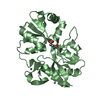
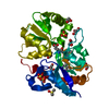
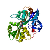
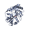
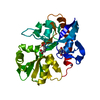
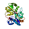
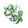
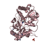
| タイトル | Crystal structure of a tetramer of GluA2 TR mutant ligand binding domains bound with glutamate at 1.26 Angstrom resolution |
|---|
 要素 要素 | Glutamate receptor 2,Glutamate receptor 2 |
|---|
 キーワード キーワード | TRANSPORT PROTEIN / tetramer / ligand-gated ion channel |
|---|
| 機能・相同性 |  機能・相同性情報 機能・相同性情報
spine synapse / dendritic spine neck / dendritic spine head / cellular response to amine stimulus / Activation of AMPA receptors / perisynaptic space / ligand-gated monoatomic cation channel activity / AMPA glutamate receptor activity / Trafficking of GluR2-containing AMPA receptors / response to lithium ion ...spine synapse / dendritic spine neck / dendritic spine head / cellular response to amine stimulus / Activation of AMPA receptors / perisynaptic space / ligand-gated monoatomic cation channel activity / AMPA glutamate receptor activity / Trafficking of GluR2-containing AMPA receptors / response to lithium ion / kainate selective glutamate receptor activity / AMPA glutamate receptor complex / cellular response to glycine / extracellularly glutamate-gated ion channel activity / ionotropic glutamate receptor complex / immunoglobulin binding / asymmetric synapse / conditioned place preference / regulation of receptor recycling / glutamate receptor binding / Unblocking of NMDA receptors, glutamate binding and activation / positive regulation of synaptic transmission / regulation of synaptic transmission, glutamatergic / response to fungicide / glutamate-gated receptor activity / cytoskeletal protein binding / regulation of long-term synaptic depression / extracellular ligand-gated monoatomic ion channel activity / cellular response to brain-derived neurotrophic factor stimulus / glutamate-gated calcium ion channel activity / presynaptic active zone membrane / somatodendritic compartment / ionotropic glutamate receptor binding / dendrite membrane / ligand-gated monoatomic ion channel activity involved in regulation of presynaptic membrane potential / dendrite cytoplasm / ionotropic glutamate receptor signaling pathway / synaptic membrane / SNARE binding / dendritic shaft / transmitter-gated monoatomic ion channel activity involved in regulation of postsynaptic membrane potential / synaptic transmission, glutamatergic / PDZ domain binding / protein tetramerization / establishment of protein localization / postsynaptic density membrane / modulation of chemical synaptic transmission / cerebral cortex development / receptor internalization / Schaffer collateral - CA1 synapse / terminal bouton / synaptic vesicle / synaptic vesicle membrane / presynapse / signaling receptor activity / amyloid-beta binding / growth cone / presynaptic membrane / scaffold protein binding / perikaryon / chemical synaptic transmission / dendritic spine / postsynaptic membrane / neuron projection / postsynaptic density / axon / external side of plasma membrane / neuronal cell body / dendrite / synapse / protein kinase binding / protein-containing complex binding / glutamatergic synapse / cell surface / endoplasmic reticulum / protein-containing complex / identical protein binding / membrane / plasma membrane類似検索 - 分子機能 Ionotropic glutamate receptor, metazoa / Ligated ion channel L-glutamate- and glycine-binding site / Ionotropic glutamate receptor, L-glutamate and glycine-binding domain / Ligated ion channel L-glutamate- and glycine-binding site / Ligand-gated ion channel / : / Ionotropic glutamate receptor / Eukaryotic homologues of bacterial periplasmic substrate binding proteins. / Receptor, ligand binding region / Receptor family ligand binding region ...Ionotropic glutamate receptor, metazoa / Ligated ion channel L-glutamate- and glycine-binding site / Ionotropic glutamate receptor, L-glutamate and glycine-binding domain / Ligated ion channel L-glutamate- and glycine-binding site / Ligand-gated ion channel / : / Ionotropic glutamate receptor / Eukaryotic homologues of bacterial periplasmic substrate binding proteins. / Receptor, ligand binding region / Receptor family ligand binding region / Periplasmic binding protein-like II / Periplasmic binding protein-like I / D-Maltodextrin-Binding Protein; domain 2 / 3-Layer(aba) Sandwich / Alpha Beta類似検索 - ドメイン・相同性 GLUTAMIC ACID / DI(HYDROXYETHYL)ETHER / PHOSPHATE ION / Glutamate receptor 2類似検索 - 構成要素 |
|---|
| 生物種 |   Rattus norvegicus (ドブネズミ) Rattus norvegicus (ドブネズミ) |
|---|
| 手法 |  X線回折 / シンクロトロン / 分子置換 / 解像度: 1.26 Å X線回折 / シンクロトロン / 分子置換 / 解像度: 1.26 Å |
|---|
 データ登録者 データ登録者 | Chebli, M. / Salazar, H. / Baranovic, J. / Carbone, A.L. / Ghisi, V. / Faelber, K. / Lau, A.Y. / Daumke, O. / Plested, A.J.R. |
|---|
 引用 引用 | ジャーナル: To Be Published
タイトル: Crystal structure of the tetrameric GluA2 ligand-binding domain in complex with glutamate at 1.26 Angstroms resolution
著者: Chebli, M. / Salazar, H. / Baranovic, J. / Carbone, A.L. / Ghisi, V. / Faelber, K. / Lau, A.Y. / Daumke, O. / Plested, A.J.R. |
|---|
| 履歴 | | 登録 | 2015年3月18日 | 登録サイト: RCSB / 処理サイト: PDBE |
|---|
| 改定 1.0 | 2016年1月13日 | Provider: repository / タイプ: Initial release |
|---|
| 改定 2.0 | 2021年3月31日 | Group: Advisory / Atomic model ...Advisory / Atomic model / Data collection / Derived calculations
カテゴリ: atom_site / atom_site_anisotrop ...atom_site / atom_site_anisotrop / pdbx_distant_solvent_atoms / pdbx_nonpoly_scheme / pdbx_struct_assembly / pdbx_struct_assembly_gen / pdbx_struct_assembly_prop / pdbx_struct_special_symmetry / pdbx_validate_close_contact / struct_site_gen
Item: _atom_site.B_iso_or_equiv / _atom_site.Cartn_x ..._atom_site.B_iso_or_equiv / _atom_site.Cartn_x / _atom_site.Cartn_y / _atom_site.Cartn_z / _atom_site.occupancy / _atom_site_anisotrop.U[1][1] / _atom_site_anisotrop.U[1][2] / _atom_site_anisotrop.U[1][3] / _atom_site_anisotrop.U[2][2] / _atom_site_anisotrop.U[2][3] / _atom_site_anisotrop.U[3][3] / _pdbx_distant_solvent_atoms.auth_seq_id / _pdbx_distant_solvent_atoms.neighbor_macromolecule_distance / _pdbx_nonpoly_scheme.auth_seq_num / _pdbx_struct_special_symmetry.auth_seq_id / _pdbx_validate_close_contact.auth_seq_id_2 / _struct_site_gen.auth_seq_id |
|---|
| 改定 2.1 | 2024年1月10日 | Group: Data collection / Database references / Refinement description
カテゴリ: chem_comp_atom / chem_comp_bond ...chem_comp_atom / chem_comp_bond / database_2 / pdbx_initial_refinement_model
Item: _database_2.pdbx_DOI / _database_2.pdbx_database_accession |
|---|
| 改定 2.2 | 2024年11月13日 | Group: Structure summary
カテゴリ: pdbx_entry_details / pdbx_modification_feature |
|---|
|
|---|
 ムービー
ムービー コントローラー


 データを開く
 基本情報
基本情報 構造の表示
構造の表示 ダウンロードとリンク
 その他のダウンロード


 PDBj
PDBj


 集合体
集合体


 タイプ: L-peptide linking / 分子量: 147.129 Da / 分子数: 2 / 由来タイプ: 合成 / 式: C5H9NO4
 分子量: 94.971 Da / 分子数: 6 / 由来タイプ: 合成 / 式: PO4
 分子量: 106.120 Da / 分子数: 4 / 由来タイプ: 合成 / 式: C4H10O3
 分子量: 18.015 Da / 分子数: 822 / 由来タイプ: 天然 / 式: H2O
 試料調製
試料調製 / ビームライン: 14.1 / 波長: 0.918 Å
 解析
解析