Monochromator: double crystal Si(111) / Protocol: SINGLE WAVELENGTH / Monochromatic (M) / Laue (L): M / Scattering type: x-ray
Radiation wavelength
Wavelength: 0.97947 Å / Relative weight: 1
Reflection
Resolution: 1.8→26.531 Å / Num. obs: 18276 / % possible obs: 99 % / Observed criterion σ(I): -3 / Redundancy: 2.58 % / Biso Wilson estimate: 22.698 Å2 / Rmerge F obs: 0.998 / Rmerge(I) obs: 0.062 / Rrim(I) all: 0.078 / Net I/σ(I): 11.04 / Num. measured all: 87402
Reflection shell
Resolution (Å)
Highest resolution (Å)
Rmerge F obs
Rmerge(I) obs
Mean I/σ(I) obs
Num. measured obs
Num. possible
Num. unique obs
Rrim(I) all
Diffraction-ID
% possible all
1.8-1.86
0.565
0.649
1.5
7957
3216
3161
0.821
1
98.3
1.86-1.94
0.757
0.458
2.2
9272
3642
3624
0.578
99.5
1.94-2.03
0.869
0.291
3.5
8848
3460
3445
0.367
99.6
2.03-2.13
0.931
0.203
4.9
8299
3226
3214
0.256
99.6
2.13-2.27
0.961
0.15
6.5
9240
3594
3583
0.189
99.7
2.27-2.44
0.974
0.119
8.3
8619
3335
3312
0.15
99.3
2.44-2.69
0.987
0.083
11.4
8874
3449
3415
0.105
99
2.69-3.07
0.994
0.056
15.8
8591
3344
3310
0.071
99
3.07-3.87
0.997
0.034
25
8937
3477
3417
0.043
98.3
3.87
0.998
0.026
31.3
8765
3452
3361
0.033
97.4
-
Phasing
Phasing
Method: SAD
-
Processing
Software
Name
Version
Classification
PDB_EXTRACT
3.1
dataextraction
SHELX
phasing
SHARP
phasing
XDS
November3, 2014BUILT=20141118
datascaling
XSCALE
datascaling
BUSTER-TNT
2.10.2
refinement
SHELXD
phasing
BUSTER
2.10.2
refinement
XDS
datareduction
Refinement
Method to determine structure: SAD / Resolution: 1.8→26.531 Å / Cor.coef. Fo:Fc: 0.9504 / Cor.coef. Fo:Fc free: 0.9404 / Occupancy max: 1 / Occupancy min: 0.4 / Cross valid method: THROUGHOUT / σ(F): 0 Details: 1. A MET-INHIBITION PROTOCOL WAS USED FOR SELENOMETHIONINE INCORPORATION DURING PROTEIN EXPRESSION. THE OCCUPANCY OF THE SE ATOMS IN THE MSE RESIDUES WAS REDUCED TO 0.75 FOR THE REDUCED ...Details: 1. A MET-INHIBITION PROTOCOL WAS USED FOR SELENOMETHIONINE INCORPORATION DURING PROTEIN EXPRESSION. THE OCCUPANCY OF THE SE ATOMS IN THE MSE RESIDUES WAS REDUCED TO 0.75 FOR THE REDUCED SCATTERING POWER DUE TO PARTIAL S-MET INCORPORATION. 2. THE MAD PHASES WERE USED AS RESTRAINTS DURING REFINEMENT. 3. MG, CL, AND GOL ARE PRESENT IN CRYSTALLIZATION/CRYO CONDITIONS. 4. ELECTRON DENSITY FOR LOOP 61-65 IS WEAK.
In the structure databanks used in Yorodumi, some data are registered as the other names, "COVID-19 virus" and "2019-nCoV". Here are the details of the virus and the list of structure data.
Jan 31, 2019. EMDB accession codes are about to change! (news from PDBe EMDB page)
EMDB accession codes are about to change! (news from PDBe EMDB page)
The allocation of 4 digits for EMDB accession codes will soon come to an end. Whilst these codes will remain in use, new EMDB accession codes will include an additional digit and will expand incrementally as the available range of codes is exhausted. The current 4-digit format prefixed with “EMD-” (i.e. EMD-XXXX) will advance to a 5-digit format (i.e. EMD-XXXXX), and so on. It is currently estimated that the 4-digit codes will be depleted around Spring 2019, at which point the 5-digit format will come into force.
The EM Navigator/Yorodumi systems omit the EMD- prefix.
Related info.:Q: What is EMD? / ID/Accession-code notation in Yorodumi/EM Navigator
Yorodumi is a browser for structure data from EMDB, PDB, SASBDB, etc.
This page is also the successor to EM Navigator detail page, and also detail information page/front-end page for Omokage search.
The word "yorodu" (or yorozu) is an old Japanese word meaning "ten thousand". "mi" (miru) is to see.
Related info.:EMDB / PDB / SASBDB / Comparison of 3 databanks / Yorodumi Search / Aug 31, 2016. New EM Navigator & Yorodumi / Yorodumi Papers / Jmol/JSmol / Function and homology information / Changes in new EM Navigator and Yorodumi
 Movie
Movie Controller

 Yorodumi
 Open data
 Basic information
 Components
 Keywords
 Function and homology information
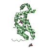
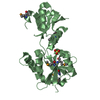
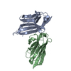
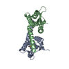
 Parabacteroides merdae ATCC 43184 (bacteria)
 X-RAY DIFFRACTION /
  Authors
 Citation
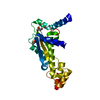
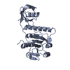
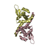
 Structure visualization
 Downloads & links
 Other downloads
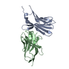


 PDBj
PDBj Assembly

 Mass: 24.305 Da / Num. of mol.: 1 / Source method: obtained synthetically / Formula: Mg
 Mass: 92.094 Da / Num. of mol.: 1 / Source method: obtained synthetically / Formula: C3H8O3
 Mass: 35.453 Da / Num. of mol.: 2 / Source method: obtained synthetically / Formula: Cl
 Mass: 18.015 Da / Num. of mol.: 174 / Source method: isolated from a natural source / Formula: H2O
 Sample preparation
 / Beamline: BL14-1 / Wavelength: 0.97947 Å
 Processing