 Movie
Movie Controller

[English] 日本語
 Yorodumi
- PDB-4yji: The Crystal Structure of a Bacterial Aryl Acylamidase Belonging t... -
+ Open data

- Basic information
| Entry | Database: PDB / ID: 4yji | ||||||
|---|---|---|---|---|---|---|---|
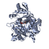
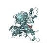
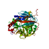
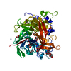
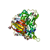
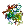
| Title | The Crystal Structure of a Bacterial Aryl Acylamidase Belonging to the Amidase signature (AS) enzymes family | ||||||
 Components Components | Aryl acylamidase | ||||||
 Keywords Keywords | HYDROLASE / amidase | ||||||
| Function / homology |  Function and homology information Function and homology information | ||||||
| Biological species |  bacterium CSBL00001 (bacteria) bacterium CSBL00001 (bacteria) | ||||||
| Method |  X-RAY DIFFRACTION / SYNCHROTRON / MOLECULAR REPLACEMENT / Resolution: 1.73 Å X-RAY DIFFRACTION / SYNCHROTRON / MOLECULAR REPLACEMENT / Resolution: 1.73 Å | ||||||
 Authors Authors | Choi, I.-G. / Lee, S. / Park, E.-H. / Ko, H.-J. / Bang, W.-G. | ||||||
 Citation Citation | Journal: Biochem.Biophys.Res.Commun. / Year: 2015 Title: Crystal structure analysis of a bacterial aryl acylamidase belonging to the amidase signature enzyme family Authors: Lee, S. / Park, E.-H. / Ko, H.-J. / Bang, W.-G. / Kim, H.Y. / Kim, K.H. / Choi, I.-G. | ||||||
| History |
|
- Structure visualization
| Structure viewer | Molecule: MolmilJmol/JSmol |
|---|
- Downloads & links
-Download
| PDBx/mmCIF format | 4yji.cif.gz | 120.1 KB | Display | PDBx/mmCIF format |
|---|---|---|---|---|
| PDB format | pdb4yji.ent.gz | 89.3 KB | Display | PDB format |
| PDBx/mmJSON format | 4yji.json.gz | Tree view | PDBx/mmJSON format | |
| Others |  Other downloads Other downloads |
-Validation report
| Arichive directory | https://data.pdbj.org/pub/pdb/validation_reports/yj/4yjiftp://data.pdbj.org/pub/pdb/validation_reports/yj/4yji | HTTPS FTP |
|---|
-Related structure data
| Related structure data | 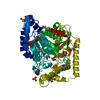 4yj6SC S: Starting model for refinement C: citing same article ( |
|---|---|
| Similar structure data |


-Links
 PDBj
PDBj
- Assembly
| Deposited unit | 
| ||||||||||||
|---|---|---|---|---|---|---|---|---|---|---|---|---|---|
| 1 |
| ||||||||||||
| Unit cell |
| ||||||||||||
| Components on special symmetry positions |
|
-Components
| #1: Protein | Mass: 53262.500 Da / Num. of mol.: 1 / Mutation: S187A Source method: isolated from a genetically manipulated source Source: (gene. exp.) bacterium CSBL00001 (bacteria) / Production host: |
|---|---|
| #2: Chemical | ChemComp-CXS /   Mass: 221.317 Da / Num. of mol.: 1 / Source method: obtained synthetically / Formula: C9H19NO3S / Comment: pH buffer*YM Mass: 221.317 Da / Num. of mol.: 1 / Source method: obtained synthetically / Formula: C9H19NO3S / Comment: pH buffer*YM |
| #3: Chemical | ChemComp-TYL /   Mass: 151.163 Da / Num. of mol.: 1 / Source method: obtained synthetically / Formula: C8H9NO2 / Comment: medication*YM Mass: 151.163 Da / Num. of mol.: 1 / Source method: obtained synthetically / Formula: C8H9NO2 / Comment: medication*YM |
| #4: Water | ChemComp-HOH /  Mass: 18.015 Da / Num. of mol.: 545 / Source method: isolated from a natural source / Formula: H2O Mass: 18.015 Da / Num. of mol.: 545 / Source method: isolated from a natural source / Formula: H2O |
-Experimental details
-Experiment
| Experiment | Method: X-RAY DIFFRACTION / Number of used crystals: 1 |
|---|
- Sample preparation
| Crystal | Density Matthews: 2.91 Å3/Da / Density % sol: 57.72 % |
|---|---|
| Crystal grow | Temperature: 295.15 K / Method: vapor diffusion, sitting drop Details: 1.2 M NaH2PO4/0.8 M K2HPO4, 0.1 M CAPS pH 10.5, 0.2 M Li2SO4 and 0.1 M ZnCl2 |
-Data collection
| Diffraction | Mean temperature: 100 K |
|---|---|
| Diffraction source | Source: SYNCHROTRON / Site: PAL/PLS  / Beamline: 5C (4A) / Wavelength: 0.979 Å / Beamline: 5C (4A) / Wavelength: 0.979 Å |
| Detector | Type: ADSC QUANTUM 315r / Detector: CCD / Date: May 22, 2014 |
| Radiation | Protocol: SINGLE WAVELENGTH / Monochromatic (M) / Laue (L): M / Scattering type: x-ray |
| Radiation wavelength | Wavelength: 0.979 Å / Relative weight: 1 |
| Reflection | Resolution: 1.73→43.84 Å / Num. obs: 65840 / % possible obs: 98.6 % / Redundancy: 26.9 % / Net I/σ(I): 11.5 |
- Processing
| Software |
| |||||||||||||||||||||||||||||||||||||||||||||||||||||||||||||||||||||||||||||||||||||||||||||||||||||||||
|---|---|---|---|---|---|---|---|---|---|---|---|---|---|---|---|---|---|---|---|---|---|---|---|---|---|---|---|---|---|---|---|---|---|---|---|---|---|---|---|---|---|---|---|---|---|---|---|---|---|---|---|---|---|---|---|---|---|---|---|---|---|---|---|---|---|---|---|---|---|---|---|---|---|---|---|---|---|---|---|---|---|---|---|---|---|---|---|---|---|---|---|---|---|---|---|---|---|---|---|---|---|---|---|---|---|---|
| Refinement | Method to determine structure: MOLECULAR REPLACEMENT Starting model: 4YJ6 Resolution: 1.73→42.97 Å / SU ML: 0.14 / Cross valid method: NONE / σ(F): 0 / Phase error: 17.43 / Stereochemistry target values: ML
| |||||||||||||||||||||||||||||||||||||||||||||||||||||||||||||||||||||||||||||||||||||||||||||||||||||||||
| Solvent computation | Shrinkage radii: 0.9 Å / VDW probe radii: 1.11 Å / Solvent model: FLAT BULK SOLVENT MODEL | |||||||||||||||||||||||||||||||||||||||||||||||||||||||||||||||||||||||||||||||||||||||||||||||||||||||||
| Refinement step | Cycle: LAST / Resolution: 1.73→42.97 Å
| |||||||||||||||||||||||||||||||||||||||||||||||||||||||||||||||||||||||||||||||||||||||||||||||||||||||||
| Refine LS restraints |
| |||||||||||||||||||||||||||||||||||||||||||||||||||||||||||||||||||||||||||||||||||||||||||||||||||||||||
| LS refinement shell |
|
