 Movie
Movie Controller

[English] 日本語
 Yorodumi
- PDB-4y9h: The 1.43 angstrom crystal structure of bacteriorhodopsin crystall... -
+ Open data

- Basic information
| Entry | Database: PDB / ID: 4y9h | |||||||||
|---|---|---|---|---|---|---|---|---|---|---|
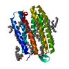
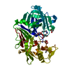
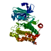
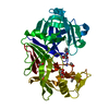
| Title | The 1.43 angstrom crystal structure of bacteriorhodopsin crystallized from bicelles | |||||||||
 Components Components | Bacteriorhodopsin | |||||||||
 Keywords Keywords | LIPID BINDING PROTEIN / bacteriorhodopsin / complex / membrane protein / Cell membrane / Multi-pass membrane protein | |||||||||
| Function / homology |  Function and homology information Function and homology informationphotoreceptor activity / phototransduction / monoatomic ion channel activity / protein domain specific binding / plasma membrane Similarity search - Function | |||||||||
| Biological species |  Halobacterium salinarum (Halophile) Halobacterium salinarum (Halophile) | |||||||||
| Method |  X-RAY DIFFRACTION / SYNCHROTRON / MOLECULAR REPLACEMENT / Resolution: 1.43 Å X-RAY DIFFRACTION / SYNCHROTRON / MOLECULAR REPLACEMENT / Resolution: 1.43 Å | |||||||||
 Authors Authors | Saiki, H. / Sugiyama, S. / Kakinouchi, K. / Kawatake, S. / Hanashima, S. / Matsumori, N. / Murata, M. | |||||||||
| Funding support |  Japan, 2items Japan, 2items
| |||||||||
 Citation Citation | Journal: To Be Published Title: The 1.43 angstrom crystal structure of bacteriorhodopsin crystallized from bicelles Authors: Saiki, H. / Sugiyama, S. / Kakinouchi, K. / Kawatake, S. / Hanashima, S. / Matsumori, N. / Murata, M. | |||||||||
| History |
|
- Structure visualization
| Structure viewer | Molecule: MolmilJmol/JSmol |
|---|
- Downloads & links
-Download
| PDBx/mmCIF format | 4y9h.cif.gz | 67.3 KB | Display | PDBx/mmCIF format |
|---|---|---|---|---|
| PDB format | pdb4y9h.ent.gz | 48.3 KB | Display | PDB format |
| PDBx/mmJSON format | 4y9h.json.gz | Tree view | PDBx/mmJSON format | |
| Others |  Other downloads Other downloads |
-Validation report
| Arichive directory | https://data.pdbj.org/pub/pdb/validation_reports/y9/4y9hftp://data.pdbj.org/pub/pdb/validation_reports/y9/4y9h | HTTPS FTP |
|---|
-Related structure data
| Similar structure data |
|---|


-Links
 PDBj
PDBj


- Assembly
| Deposited unit | 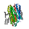
| ||||||||
|---|---|---|---|---|---|---|---|---|---|
| 1 |
| ||||||||
| Unit cell |
| ||||||||
| Components on special symmetry positions |
|
-Components
-Protein , 1 types, 1 molecules A
| #1: Protein | Mass: 24760.316 Da / Num. of mol.: 1 / Source method: isolated from a natural source Source: (natural) Halobacterium salinarum (strain ATCC 29341 / DSM 671 / R1) (Halophile)Strain: ATCC 29341 / DSM 671 / R1 / References: UniProt: B0R5N9 |
|---|
-Non-polymers , 8 types, 111 molecules 


| #2: Chemical | ChemComp-RET /  Mass: 284.436 Da / Num. of mol.: 1 / Source method: obtained synthetically / Formula: C20H28O Mass: 284.436 Da / Num. of mol.: 1 / Source method: obtained synthetically / Formula: C20H28O | ||||||||
|---|---|---|---|---|---|---|---|---|---|
| #3: Chemical | ChemComp-PO4 /  Mass: 94.971 Da / Num. of mol.: 1 / Source method: obtained synthetically / Formula: PO4 Mass: 94.971 Da / Num. of mol.: 1 / Source method: obtained synthetically / Formula: PO4 | ||||||||
| #4: Chemical | ChemComp-HP6 /  Mass: 100.202 Da / Num. of mol.: 1 / Source method: obtained synthetically / Formula: C7H16 Mass: 100.202 Da / Num. of mol.: 1 / Source method: obtained synthetically / Formula: C7H16 | ||||||||
| #5: Chemical | ChemComp-D12 /  Mass: 170.335 Da / Num. of mol.: 8 / Source method: obtained synthetically / Formula: C12H26 Mass: 170.335 Da / Num. of mol.: 8 / Source method: obtained synthetically / Formula: C12H26#6: Chemical | ChemComp-D10 /  Mass: 142.282 Da / Num. of mol.: 4 / Source method: obtained synthetically / Formula: C10H22 Mass: 142.282 Da / Num. of mol.: 4 / Source method: obtained synthetically / Formula: C10H22#7: Chemical | ChemComp-R16 / |  Mass: 226.441 Da / Num. of mol.: 1 / Source method: obtained synthetically / Formula: C16H34 Mass: 226.441 Da / Num. of mol.: 1 / Source method: obtained synthetically / Formula: C16H34#8: Chemical | ChemComp-DD9 / |  Mass: 128.255 Da / Num. of mol.: 1 / Source method: obtained synthetically / Formula: C9H20 Mass: 128.255 Da / Num. of mol.: 1 / Source method: obtained synthetically / Formula: C9H20#9: Water | ChemComp-HOH / | Mass: 18.015 Da / Num. of mol.: 94 / Source method: isolated from a natural source / Formula: H2O |
-Experimental details
-Experiment
| Experiment | Method: X-RAY DIFFRACTION |
|---|
- Sample preparation
| Crystal | Density Matthews: 3.01 Å3/Da / Density % sol: 59.15 % |
|---|---|
| Crystal grow | Temperature: 293 K / Method: vapor diffusion, sitting drop / pH: 4.2 Details: 180mM 1,6-hexanediol, 2.6M NaH2PO4, 3.5% triethyleneglycol, pH4.2 |
-Data collection
| Diffraction | Mean temperature: 100 K |
|---|---|
| Diffraction source | Source: SYNCHROTRON / Site: SPring-8 / Beamline: BL44XU / Wavelength: 0.9 Å |
| Detector | Type: RAYONIX MX300HE / Detector: CCD / Date: Jan 20, 2015 |
| Radiation | Protocol: SINGLE WAVELENGTH / Monochromatic (M) / Laue (L): M / Scattering type: x-ray |
| Radiation wavelength | Wavelength: 0.9 Å / Relative weight: 1 |
| Reflection | Resolution: 1.43→50 Å / Num. obs: 55216 / % possible obs: 99.4 % / Redundancy: 12.5 % / Rmerge(I) obs: 0.13 / Net I/σ(I): 6.4 |
| Reflection shell | Resolution: 1.43→1.45 Å / Redundancy: 9.3 % / Rmerge(I) obs: 0.8 / Mean I/σ(I) obs: 2.1 / % possible all: 98.5 |
- Processing
| Software |
| ||||||||||||||||||||||||||||||||||||||||||||||||||||||||||||||||||||||||||||||||||||||||||||||||||||||||||||||||||||||||||||||||||||||||||||||||||||||||||||||||||||||||||||||||||||||
|---|---|---|---|---|---|---|---|---|---|---|---|---|---|---|---|---|---|---|---|---|---|---|---|---|---|---|---|---|---|---|---|---|---|---|---|---|---|---|---|---|---|---|---|---|---|---|---|---|---|---|---|---|---|---|---|---|---|---|---|---|---|---|---|---|---|---|---|---|---|---|---|---|---|---|---|---|---|---|---|---|---|---|---|---|---|---|---|---|---|---|---|---|---|---|---|---|---|---|---|---|---|---|---|---|---|---|---|---|---|---|---|---|---|---|---|---|---|---|---|---|---|---|---|---|---|---|---|---|---|---|---|---|---|---|---|---|---|---|---|---|---|---|---|---|---|---|---|---|---|---|---|---|---|---|---|---|---|---|---|---|---|---|---|---|---|---|---|---|---|---|---|---|---|---|---|---|---|---|---|---|---|---|---|
| Refinement | Method to determine structure: MOLECULAR REPLACEMENT / Resolution: 1.43→50 Å / Cor.coef. Fo:Fc: 0.96 / Cor.coef. Fo:Fc free: 0.951 / SU B: 1.675 / SU ML: 0.056 / Cross valid method: THROUGHOUT / ESU R: 0.064 / ESU R Free: 0.065 / Stereochemistry target values: MAXIMUM LIKELIHOOD / Details: HYDROGENS HAVE BEEN ADDED IN THE RIDING POSITIONS
| ||||||||||||||||||||||||||||||||||||||||||||||||||||||||||||||||||||||||||||||||||||||||||||||||||||||||||||||||||||||||||||||||||||||||||||||||||||||||||||||||||||||||||||||||||||||
| Solvent computation | Ion probe radii: 0.8 Å / Shrinkage radii: 0.8 Å / VDW probe radii: 1.2 Å / Solvent model: MASK | ||||||||||||||||||||||||||||||||||||||||||||||||||||||||||||||||||||||||||||||||||||||||||||||||||||||||||||||||||||||||||||||||||||||||||||||||||||||||||||||||||||||||||||||||||||||
| Displacement parameters | Biso mean: 25.844 Å2
| ||||||||||||||||||||||||||||||||||||||||||||||||||||||||||||||||||||||||||||||||||||||||||||||||||||||||||||||||||||||||||||||||||||||||||||||||||||||||||||||||||||||||||||||||||||||
| Refinement step | Cycle: LAST / Resolution: 1.43→50 Å
| ||||||||||||||||||||||||||||||||||||||||||||||||||||||||||||||||||||||||||||||||||||||||||||||||||||||||||||||||||||||||||||||||||||||||||||||||||||||||||||||||||||||||||||||||||||||
| Refine LS restraints |
|