 Movie
Movie Controller


+ Open data

- Basic information
| Entry | Database: PDB / ID: 4y7p | ||||||
|---|---|---|---|---|---|---|---|
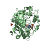
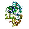
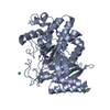
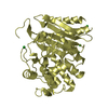
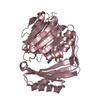
| Title | Structure of alkaline D-peptidase from Bacillus cereus | ||||||
 Components Components | Alkaline D-peptidase | ||||||
 Keywords Keywords | HYDROLASE / Penicillin binding protein / apo form | ||||||
| Function / homology |  Function and homology information Function and homology information: / Beta-lactamase-related / Beta-lactamase / Beta-lactamase / DD-peptidase/beta-lactamase superfamily / Beta-lactamase/transpeptidase-like / 3-Layer(aba) Sandwich / Alpha Beta Similarity search - Domain/homology | ||||||
| Biological species |  | ||||||
| Method |  X-RAY DIFFRACTION / MOLECULAR REPLACEMENT / Resolution: 2.1 Å X-RAY DIFFRACTION / MOLECULAR REPLACEMENT / Resolution: 2.1 Å | ||||||
 Authors Authors | Nakano, S. / Okazaki, S. / Ishitsubo, E. / Kawahara, N. / Komeda, H. / Tokiwa, H. / Asano, Y. | ||||||
 Citation Citation | Journal: Sci Rep / Year: 2015 Title: Structural and computational analysis of peptide recognition mechanism of class-C type penicillin binding protein, alkaline D-peptidase from Bacillus cereus DF4-B Authors: Nakano, S. / Okazaki, S. / Ishitsubo, E. / Kawahara, N. / Komeda, H. / Tokiwa, H. / Asano, Y. | ||||||
| History |
|
- Structure visualization
| Structure viewer | Molecule: MolmilJmol/JSmol |
|---|
- Downloads & links
-Download
| PDBx/mmCIF format | 4y7p.cif.gz | 86.9 KB | Display | PDBx/mmCIF format |
|---|---|---|---|---|
| PDB format | pdb4y7p.ent.gz | 62.8 KB | Display | PDB format |
| PDBx/mmJSON format | 4y7p.json.gz | Tree view | PDBx/mmJSON format | |
| Others |  Other downloads Other downloads |
-Validation report
| Summary document | 4y7p_validation.pdf.gz | 438 KB | Display | wwPDB validaton report |
|---|---|---|---|---|
| Full document | 4y7p_full_validation.pdf.gz | 443.9 KB | Display | |
| Data in XML | 4y7p_validation.xml.gz | 16.8 KB | Display | |
| Data in CIF | 4y7p_validation.cif.gz | 24.5 KB | Display | |
| Arichive directory | https://data.pdbj.org/pub/pdb/validation_reports/y7/4y7pftp://data.pdbj.org/pub/pdb/validation_reports/y7/4y7p | HTTPS FTP |
-Related structure data
| Related structure data |  1hvbS S: Starting model for refinement |
|---|---|
| Similar structure data |


-Links
 PDBj
PDBj

- Assembly
| Deposited unit | 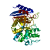
| ||||||||
|---|---|---|---|---|---|---|---|---|---|
| 1 |
| ||||||||
| 2 |
| ||||||||
| Unit cell |
|
-Components
| #1: Protein | Mass: 42078.578 Da / Num. of mol.: 1 Source method: isolated from a genetically manipulated source Source: (gene. exp.) | ||
|---|---|---|---|
| #2: Chemical |   Mass: 58.082 Da / Num. of mol.: 3 / Source method: obtained synthetically / Formula: CNS Mass: 58.082 Da / Num. of mol.: 3 / Source method: obtained synthetically / Formula: CNS#3: Water | ChemComp-HOH / |  Mass: 18.015 Da / Num. of mol.: 203 / Source method: isolated from a natural source / Formula: H2O Mass: 18.015 Da / Num. of mol.: 203 / Source method: isolated from a natural source / Formula: H2O |
-Experimental details
-Experiment
| Experiment | Method: X-RAY DIFFRACTION |
|---|
- Sample preparation
| Crystal | Density Matthews: 3.66 Å3/Da / Density % sol: 66.37 % |
|---|---|
| Crystal grow | Temperature: 277 K / Method: vapor diffusion, hanging drop / pH: 7 / Details: 20% PEG3350, 0.2M sodium thiocyanate, 3% D-glucose |
-Data collection
| Diffraction | Mean temperature: 100 K |
|---|---|
| Diffraction source | Source: ROTATING ANODE / Type: RIGAKU MICROMAX-007 / Wavelength: 1.5418 Å |
| Detector | Type: RIGAKU RAXIS VII / Detector: IMAGE PLATE / Date: Jun 14, 2013 |
| Radiation | Protocol: SINGLE WAVELENGTH / Monochromatic (M) / Laue (L): M / Scattering type: x-ray |
| Radiation wavelength | Wavelength: 1.5418 Å / Relative weight: 1 |
| Reflection | Resolution: 2.1→100 Å / Num. obs: 69524 / % possible obs: 99.9 % / Redundancy: 4.4 % / Rmerge(I) obs: 0.068 / Net I/σ(I): 33.3 |
| Reflection shell | Resolution: 2.1→2.14 Å / Rmerge(I) obs: 0.487 / Num. unique all: 69524 |
- Processing
| Software |
| ||||||||||||||||||||||||||||||||||||||||||||||||||||||||||||||||||||||||||||||||||||||||||||||||||||||||||||||||||||||||||||||||||||||||||||||||||||||||||||||||||||||||||||||||||||||
|---|---|---|---|---|---|---|---|---|---|---|---|---|---|---|---|---|---|---|---|---|---|---|---|---|---|---|---|---|---|---|---|---|---|---|---|---|---|---|---|---|---|---|---|---|---|---|---|---|---|---|---|---|---|---|---|---|---|---|---|---|---|---|---|---|---|---|---|---|---|---|---|---|---|---|---|---|---|---|---|---|---|---|---|---|---|---|---|---|---|---|---|---|---|---|---|---|---|---|---|---|---|---|---|---|---|---|---|---|---|---|---|---|---|---|---|---|---|---|---|---|---|---|---|---|---|---|---|---|---|---|---|---|---|---|---|---|---|---|---|---|---|---|---|---|---|---|---|---|---|---|---|---|---|---|---|---|---|---|---|---|---|---|---|---|---|---|---|---|---|---|---|---|---|---|---|---|---|---|---|---|---|---|---|
| Refinement | Method to determine structure: MOLECULAR REPLACEMENT Starting model: 1HVB Resolution: 2.1→24.22 Å / Cor.coef. Fo:Fc: 0.962 / Cor.coef. Fo:Fc free: 0.943 / SU B: 3.234 / SU ML: 0.084 / Cross valid method: THROUGHOUT / ESU R: 0.131 / ESU R Free: 0.135 / Stereochemistry target values: MAXIMUM LIKELIHOOD / Details: HYDROGENS HAVE BEEN ADDED IN THE RIDING POSITIONS
| ||||||||||||||||||||||||||||||||||||||||||||||||||||||||||||||||||||||||||||||||||||||||||||||||||||||||||||||||||||||||||||||||||||||||||||||||||||||||||||||||||||||||||||||||||||||
| Solvent computation | Ion probe radii: 0.8 Å / Shrinkage radii: 0.8 Å / VDW probe radii: 1.2 Å / Solvent model: MASK | ||||||||||||||||||||||||||||||||||||||||||||||||||||||||||||||||||||||||||||||||||||||||||||||||||||||||||||||||||||||||||||||||||||||||||||||||||||||||||||||||||||||||||||||||||||||
| Displacement parameters | Biso mean: 34.605 Å2
| ||||||||||||||||||||||||||||||||||||||||||||||||||||||||||||||||||||||||||||||||||||||||||||||||||||||||||||||||||||||||||||||||||||||||||||||||||||||||||||||||||||||||||||||||||||||
| Refinement step | Cycle: 1 / Resolution: 2.1→24.22 Å
| ||||||||||||||||||||||||||||||||||||||||||||||||||||||||||||||||||||||||||||||||||||||||||||||||||||||||||||||||||||||||||||||||||||||||||||||||||||||||||||||||||||||||||||||||||||||
| Refine LS restraints |
|