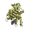
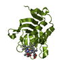
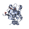
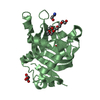
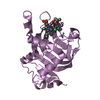
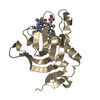
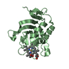
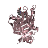
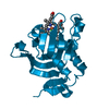
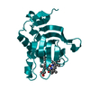
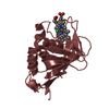
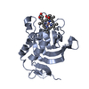
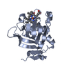
登録情報 データベース : PDB / ID : 4xzdタイトル Crystal Structure of Wild-type HasA from Yersinia pseudotuberculosis Extracellular heme acquisition hemophore HasA キーワード / / 機能・相同性 分子機能 ドメイン・相同性 構成要素
/ / / / / / / 生物種 Yersinia pseudotuberculosis IP 32953 (仮性結核菌)手法 / / / 解像度 : 1.7 Å データ登録者 Kanadani, M. / Hino, T. / Nagano, S. / Sato, T. / Ozaki, S. 資金援助 組織 認可番号 国 MEXT 25291015 MEXT 25410176
ジャーナル : J.Inorg.Biochem. / 年 : 2015タイトル : The crystal structure of heme acquisition system A from Yersinia pseudotuberculosis (HasAypt): Roles of the axial ligand Tyr75 and two distal arginines in heme binding著者 : Kanadani, M. / Sato, T. / Hino, T. / Nagano, S. / Ozaki, S. 履歴 登録 2015年2月4日 登録サイト / 処理サイト 改定 1.0 2015年8月12日 Provider / タイプ 改定 1.1 2016年1月6日 Group 改定 1.2 2020年2月5日 Group / Database references / Derived calculationsカテゴリ / diffrn_source / pdbx_struct_oper_listItem / _diffrn_source.pdbx_synchrotron_site / _pdbx_struct_oper_list.symmetry_operation改定 1.3 2023年11月8日 Group Data collection / Database references ... Data collection / Database references / Derived calculations / Refinement description カテゴリ chem_comp_atom / chem_comp_bond ... chem_comp_atom / chem_comp_bond / database_2 / pdbx_initial_refinement_model / struct_conn Item _database_2.pdbx_DOI / _database_2.pdbx_database_accession ... _database_2.pdbx_DOI / _database_2.pdbx_database_accession / _struct_conn.pdbx_dist_value / _struct_conn.ptnr1_auth_asym_id / _struct_conn.ptnr1_auth_comp_id / _struct_conn.ptnr1_auth_seq_id / _struct_conn.ptnr1_label_asym_id / _struct_conn.ptnr1_label_atom_id / _struct_conn.ptnr1_label_comp_id / _struct_conn.ptnr1_label_seq_id / _struct_conn.ptnr2_auth_asym_id / _struct_conn.ptnr2_auth_comp_id / _struct_conn.ptnr2_auth_seq_id / _struct_conn.ptnr2_label_asym_id / _struct_conn.ptnr2_label_atom_id / _struct_conn.ptnr2_label_comp_id
すべて表示 表示を減らす
 ムービー
ムービー コントローラー


 データを開く
 基本情報
基本情報 要素
要素 キーワード
キーワード 機能・相同性情報
 Yersinia pseudotuberculosis IP 32953 (仮性結核菌)
 X線回折 /
  データ登録者
 日本, 2件
  引用
引用 構造の表示
構造の表示 ダウンロードとリンク
 その他のダウンロード


 PDBj
PDBj 集合体
集合体
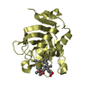

 分子量: 616.487 Da / 分子数: 2 / 由来タイプ: 合成 / 式: C34H32FeN4O4
 分子量: 18.015 Da / 分子数: 551 / 由来タイプ: 天然 / 式: H2O
 試料調製
試料調製 解析
解析