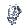
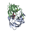
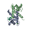
[English] 日本語
Yorodumi
- PDB-4xyn: X-ray structure of Ca(2+)-S100B with human RAGE-derived W61 peptide -

+
Open data


ID or keywords:

Loading...

-
Basic information

Entry
Database: PDB / ID: 4xyn
TitleX-ray structure of Ca(2+)-S100B with human RAGE-derived W61 peptide
Components
  • Protein S100-B
  • Receptor for advanced glycation endproducts-derived peptide (W61)
KeywordsMembrane protein / metal binding protein
Function / homology
Function and homology information


regulation of CD4-positive, alpha-beta T cell activation / negative regulation of blood circulation / advanced glycation end-product receptor activity / positive regulation of endothelin production / regulation of T cell mediated cytotoxicity / glucose mediated signaling pathway / negative regulation of long-term synaptic depression / positive regulation of monocyte extravasation / positive regulation of aspartic-type endopeptidase activity involved in amyloid precursor protein catabolic process / positive regulation of dendritic cell differentiation ...regulation of CD4-positive, alpha-beta T cell activation / negative regulation of blood circulation / advanced glycation end-product receptor activity / positive regulation of endothelin production / regulation of T cell mediated cytotoxicity / glucose mediated signaling pathway / negative regulation of long-term synaptic depression / positive regulation of monocyte extravasation / positive regulation of aspartic-type endopeptidase activity involved in amyloid precursor protein catabolic process / positive regulation of dendritic cell differentiation / adaptive thermogenesis / regulation of p38MAPK cascade / regulation of non-canonical NF-kappaB signal transduction / sympathetic neuron projection extension / RAGE receptor binding / scavenger receptor activity / induction of positive chemotaxis / ion binding / transcytosis / protein localization to membrane / positive regulation of monocyte chemotactic protein-1 production / positive regulation of heterotypic cell-cell adhesion / S100 protein binding / regulation of long-term synaptic potentiation / regulation of spontaneous synaptic transmission / laminin receptor activity / positive regulation of p38MAPK cascade / negative regulation of interleukin-10 production / positive regulation of activated T cell proliferation / regulation of neuronal synaptic plasticity / response to amyloid-beta / TRAF6 mediated NF-kB activation / Advanced glycosylation endproduct receptor signaling / transport across blood-brain barrier / negative regulation of long-term synaptic potentiation / positive regulation of chemokine production / Nuclear signaling by ERBB4 / ruffle / positive regulation of interleukin-12 production / positive regulation of neuron differentiation / axonogenesis / positive regulation of interleukin-1 beta production / sarcoplasmic reticulum / central nervous system development / astrocyte activation / positive regulation of JNK cascade / microglial cell activation / TAK1-dependent IKK and NF-kappa-B activation / regulation of synaptic plasticity / tau protein binding / memory / fibrillar center / response to wounding / positive regulation of non-canonical NF-kappaB signal transduction / cellular response to amyloid-beta / transmembrane signaling receptor activity / calcium-dependent protein binding / positive regulation of interleukin-6 production / positive regulation of tumor necrosis factor production / neuron projection development / cell junction / signaling receptor activity / positive regulation of NF-kappaB transcription factor activity / amyloid-beta binding / regulation of inflammatory response / postsynapse / positive regulation of canonical NF-kappaB signal transduction / molecular adaptor activity / learning or memory / cell surface receptor signaling pathway / positive regulation of ERK1 and ERK2 cascade / cell adhesion / response to hypoxia / inflammatory response / positive regulation of protein phosphorylation / apical plasma membrane / intracellular membrane-bounded organelle / neuronal cell body / calcium ion binding / positive regulation of cell population proliferation / protein-containing complex binding / perinuclear region of cytoplasm / cell surface / protein homodimerization activity / extracellular space / zinc ion binding / extracellular region / nucleoplasm / identical protein binding / nucleus / plasma membrane / cytoplasm / cytosol
Similarity search - Function
Protein S100-B / S-100/ICaBP type calcium binding protein signature. / S100/Calcium binding protein 7/8-like, conserved site / S100/CaBP-9k-type, calcium binding, subdomain / S-100/ICaBP type calcium binding domain / S-100/ICaBP type calcium binding domain / CD80-like, immunoglobulin C2-set / CD80-like C2-set immunoglobulin domain / EF hand / Immunoglobulin ...Protein S100-B / S-100/ICaBP type calcium binding protein signature. / S100/Calcium binding protein 7/8-like, conserved site / S100/CaBP-9k-type, calcium binding, subdomain / S-100/ICaBP type calcium binding domain / S-100/ICaBP type calcium binding domain / CD80-like, immunoglobulin C2-set / CD80-like C2-set immunoglobulin domain / EF hand / Immunoglobulin / Immunoglobulin domain / Immunoglobulin domain / EF-hand / Recoverin; domain 1 / Immunoglobulin subtype 2 / Immunoglobulin C-2 Type / EF-hand, calcium binding motif / EF-Hand 1, calcium-binding site / EF-hand calcium-binding domain. / EF-hand calcium-binding domain profile. / EF-hand domain / EF-hand domain pair / Immunoglobulin/major histocompatibility complex, conserved site / Immunoglobulin subtype / Immunoglobulin / Immunoglobulins and major histocompatibility complex proteins signature. / Ig-like domain profile. / Immunoglobulin-like domain / Immunoglobulin-like domain superfamily / Immunoglobulin-like fold / Orthogonal Bundle / Mainly Alpha
Similarity search - Domain/homology
Protein S100-B / Advanced glycosylation end product-specific receptor
Similarity search - Component
Biological speciesHomo sapiens (human)
synthetic construct (others)
MethodX-RAY DIFFRACTION / SYNCHROTRON / MOLECULAR REPLACEMENT / Resolution: 2.55 Å
AuthorsJensen, J.L. / Indurthi, V.S.K. / Neau, D. / Vetter, S.W. / Colbert, C.L.
CitationJournal: Acta Crystallogr.,Sect.D / Year: 2015
Title: Structural insights into the binding of the human receptor for advanced glycation end products (RAGE) by S100B, as revealed by an S100B-RAGE-derived peptide complex.
Authors: Jensen, J.L. / Indurthi, V.S. / Neau, D.B. / Vetter, S.W. / Colbert, C.L.
History
DepositionFeb 2, 2015Deposition site: RCSB / Processing site: RCSB
SupersessionMay 13, 2015ID: 4N6I
Revision 1.0May 13, 2015Provider: repository / Type: Initial release
Revision 1.1May 20, 2015Group: Database references
Revision 1.2May 27, 2015Group: Data collection
Revision 1.3Nov 22, 2017Group: Advisory / Derived calculations ...Advisory / Derived calculations / Refinement description / Source and taxonomy
Category: entity_src_gen / pdbx_entity_src_syn ...entity_src_gen / pdbx_entity_src_syn / pdbx_struct_oper_list / pdbx_unobs_or_zero_occ_atoms / pdbx_validate_close_contact / software
Item: _entity_src_gen.pdbx_alt_source_flag / _pdbx_entity_src_syn.pdbx_alt_source_flag ..._entity_src_gen.pdbx_alt_source_flag / _pdbx_entity_src_syn.pdbx_alt_source_flag / _pdbx_struct_oper_list.symmetry_operation / _software.classification / _software.name
Revision 1.4Feb 28, 2024Group: Advisory / Data collection ...Advisory / Data collection / Database references / Derived calculations / Refinement description
Category: chem_comp_atom / chem_comp_bond ...chem_comp_atom / chem_comp_bond / database_2 / pdbx_unobs_or_zero_occ_atoms / software / struct_conn
Item: _database_2.pdbx_DOI / _database_2.pdbx_database_accession ..._database_2.pdbx_DOI / _database_2.pdbx_database_accession / _software.name / _struct_conn.pdbx_dist_value / _struct_conn.ptnr1_auth_asym_id / _struct_conn.ptnr1_auth_comp_id / _struct_conn.ptnr1_auth_seq_id / _struct_conn.ptnr1_label_asym_id / _struct_conn.ptnr1_label_atom_id / _struct_conn.ptnr1_label_comp_id / _struct_conn.ptnr1_label_seq_id / _struct_conn.ptnr2_auth_asym_id / _struct_conn.ptnr2_auth_comp_id / _struct_conn.ptnr2_auth_seq_id / _struct_conn.ptnr2_label_asym_id / _struct_conn.ptnr2_label_atom_id / _struct_conn.ptnr2_label_comp_id

-
Structure visualization

Structure viewerMolecule:
MolmilJmol/JSmol

Downloads & links

-
Assembly

Deposited unit
P: Receptor for advanced glycation endproducts-derived peptide (W61)
A: Protein S100-B
B: Protein S100-B
C: Protein S100-B
D: Protein S100-B
hetero molecules


Theoretical massNumber of molelcules
Total (without water)44,87513
Polymers44,5545
Non-polymers3218
Water63135
1
P: Receptor for advanced glycation endproducts-derived peptide (W61)
A: Protein S100-B
B: Protein S100-B
hetero molecules


Theoretical massNumber of molelcules
Total (without water)23,2607
Polymers23,1003
Non-polymers1604
Water543
TypeNameSymmetry operationNumber
identity operation1_555x,y,z1
Buried area4260 Å2
ΔGint-92 kcal/mol
Surface area10000 Å2
MethodPISA
2
C: Protein S100-B
D: Protein S100-B
hetero molecules


Theoretical massNumber of molelcules
Total (without water)21,6146
Polymers21,4542
Non-polymers1604
Water362
TypeNameSymmetry operationNumber
identity operation1_555x,y,z1
Buried area3060 Å2
ΔGint-70 kcal/mol
Surface area9550 Å2
MethodPISA
Unit cell
Length a, b, c (Å)68.785, 86.684, 66.534
Angle α, β, γ (deg.)90.00, 90.00, 90.00
Int Tables number18
Space group name H-MP21212

-
Components

#1: Protein/peptide Receptor for advanced glycation endproducts-derived peptide (W61)


Mass: 1645.814 Da / Num. of mol.: 1 / Source method: obtained synthetically / Source: (synth.) synthetic construct (others) / References: UniProt: Q15109*PLUS
#2: Protein
Protein S100-B / S-100 protein beta chain / S-100 protein subunit beta / S100 calcium-binding protein B


Mass: 10727.037 Da / Num. of mol.: 4
Source method: isolated from a genetically manipulated source
Source: (gene. exp.) Homo sapiens (human) / Gene: S100B / Production host: Escherichia coli (E. coli) / References: UniProt: P04271
#3: Chemical
ChemComp-CA / CALCIUM ION


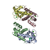
Mass: 40.078 Da / Num. of mol.: 8 / Source method: obtained synthetically / Formula: Ca
#4: Water ChemComp-HOH / water


Mass: 18.015 Da / Num. of mol.: 35 / Source method: isolated from a natural source / Formula: H2O

-
Experimental details

-
Experiment

ExperimentMethod: X-RAY DIFFRACTION

-
Sample preparation

CrystalDensity Matthews: 2.4 Å3/Da / Density % sol: 48.7 %
Crystal growTemperature: 293 K / Method: vapor diffusion, sitting drop / pH: 6.8
Details: 0.1M Na cacodylate, pH 6.8, 25% w/v PEG 3350, 9mM CaCl2

-
Data collection

DiffractionMean temperature: 100 K
Diffraction sourceSource: SYNCHROTRON / Site: APS / Beamline: 24-ID-C / Wavelength: 0.9792 Å
DetectorType: DECTRIS PILATUS 6M-F / Detector: PIXEL / Date: Nov 5, 2012
RadiationMonochromator: SI(111) / Protocol: SINGLE WAVELENGTH / Monochromatic (M) / Laue (L): M / Scattering type: x-ray
Radiation wavelengthWavelength: 0.9792 Å / Relative weight: 1
ReflectionResolution: 2.55→53.882 Å / Num. all: 35655 / Num. obs: 12927 / % possible obs: 95.7 % / Redundancy: 2.8 % / Net I/σ(I): 12.9
Reflection shellResolution: 2.55→2.64 Å / Mean I/σ(I) obs: 2.2 / % possible all: 98.8

-
Processing

Software
NameVersionClassification
PHENIX(phenix.refine: 1.8.4_1496)refinement
ADSCdata collection
XDSdata scaling
autoPROCdata scaling
Aimlessdata scaling
Cootmodel building
BALBESphasing
RefinementMethod to determine structure: MOLECULAR REPLACEMENT / Resolution: 2.55→53.882 Å / SU ML: 0.41 / Cross valid method: FREE R-VALUE / σ(F): 1.35 / Phase error: 27.87 / Stereochemistry target values: ML
RfactorNum. reflection% reflection
Rfree0.2537 639 4.94 %
Rwork0.2026 --
obs0.2053 12927 95.64 %
Solvent computationShrinkage radii: 0.9 Å / VDW probe radii: 1.11 Å / Solvent model: FLAT BULK SOLVENT MODEL
Refinement stepCycle: LAST / Resolution: 2.55→53.882 Å
ProteinNucleic acidLigandSolventTotal
Num. atoms2992 0 8 35 3035
Refine LS restraints
Refine-IDTypeDev idealNumber
X-RAY DIFFRACTIONf_bond_d0.0153051
X-RAY DIFFRACTIONf_angle_d1.54081
X-RAY DIFFRACTIONf_dihedral_angle_d14.1321148
X-RAY DIFFRACTIONf_chiral_restr0.066442
X-RAY DIFFRACTIONf_plane_restr0.008534
LS refinement shell
Resolution (Å)Rfactor RfreeNum. reflection RfreeRfactor RworkNum. reflection RworkRefine-ID% reflection obs (%)
2.55-2.74580.36511240.27792492X-RAY DIFFRACTION98
2.7458-3.02210.32821300.24952428X-RAY DIFFRACTION97
3.0221-3.45940.28971260.23512468X-RAY DIFFRACTION97
3.4594-4.35820.21971370.17422456X-RAY DIFFRACTION96
4.3582-53.89410.22441220.17962444X-RAY DIFFRACTION90

+
About Yorodumi

-
News

-
Feb 9, 2022. New format data for meta-information of EMDB entries

New format data for meta-information of EMDB entries

  • Version 3 of the EMDB header file is now the official format.
  • The previous official version 1.9 will be removed from the archive.

Related info.:EMDB header

External links:wwPDB to switch to version 3 of the EMDB data model

-
Aug 12, 2020. Covid-19 info

Covid-19 info

URL: https://pdbjlvh1.pdbj.org/emnavi/covid19.php

New page: Covid-19 featured information page in EM Navigator.

Related info.:Covid-19 info / Mar 5, 2020. Novel coronavirus structure data

+
Mar 5, 2020. Novel coronavirus structure data

Novel coronavirus structure data

Related info.:Yorodumi Speices / Aug 12, 2020. Covid-19 info

External links:COVID-19 featured content - PDBj / Molecule of the Month (242):Coronavirus Proteases

+
Jan 31, 2019. EMDB accession codes are about to change! (news from PDBe EMDB page)

EMDB accession codes are about to change! (news from PDBe EMDB page)

  • The allocation of 4 digits for EMDB accession codes will soon come to an end. Whilst these codes will remain in use, new EMDB accession codes will include an additional digit and will expand incrementally as the available range of codes is exhausted. The current 4-digit format prefixed with “EMD-” (i.e. EMD-XXXX) will advance to a 5-digit format (i.e. EMD-XXXXX), and so on. It is currently estimated that the 4-digit codes will be depleted around Spring 2019, at which point the 5-digit format will come into force.
  • The EM Navigator/Yorodumi systems omit the EMD- prefix.

Related info.:Q: What is EMD? / ID/Accession-code notation in Yorodumi/EM Navigator

External links:EMDB Accession Codes are Changing Soon! / Contact to PDBj

+
Jul 12, 2017. Major update of PDB

Major update of PDB

  • wwPDB released updated PDB data conforming to the new PDBx/mmCIF dictionary.
  • This is a major update changing the version number from 4 to 5, and with Remediation, in which all the entries are updated.
  • In this update, many items about electron microscopy experimental information are reorganized (e.g. em_software).
  • Now, EM Navigator and Yorodumi are based on the updated data.

External links:wwPDB Remediation / Enriched Model Files Conforming to OneDep Data Standards Now Available in the PDB FTP Archive

-
Yorodumi

Thousand views of thousand structures

  • Yorodumi is a browser for structure data from EMDB, PDB, SASBDB, etc.
  • This page is also the successor to EM Navigator detail page, and also detail information page/front-end page for Omokage search.
  • The word "yorodu" (or yorozu) is an old Japanese word meaning "ten thousand". "mi" (miru) is to see.

Related info.:EMDB / PDB / SASBDB / Comparison of 3 databanks / Yorodumi Search / Aug 31, 2016. New EM Navigator & Yorodumi / Yorodumi Papers / Jmol/JSmol / Function and homology information / Changes in new EM Navigator and Yorodumi

Read more