 Movie
Movie Controller

[English] 日本語
 Yorodumi
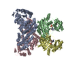
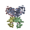
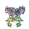
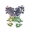
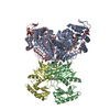
- PDB-4xym: Ca. Korarchaeum cryptofilum dinucleotide forming Acetyl-coenzyme ... -
+ Open data

- Basic information
| Entry | Database: PDB / ID: 4xym | ||||||
|---|---|---|---|---|---|---|---|
| Title | Ca. Korarchaeum cryptofilum dinucleotide forming Acetyl-coenzyme A synthetase 1 in complex with coenzyme A, Ca-AMPCP and HgCl+ | ||||||
 Components Components |
| ||||||
 Keywords Keywords | LIGASE / Dinucleotide forming acetyl-CoA synthetase / complex / ACD | ||||||
| Function / homology |  Function and homology information Function and homology informationacetate-CoA ligase (ADP-forming) activity / ligase activity / protein-lysine-acetyltransferase activity / nucleotide binding / ATP binding / metal ion binding Similarity search - Function | ||||||
| Biological species | Korarchaeum cryptofilum | ||||||
| Method |  X-RAY DIFFRACTION / SYNCHROTRON / MOLECULAR REPLACEMENT / Resolution: 1.9 Å X-RAY DIFFRACTION / SYNCHROTRON / MOLECULAR REPLACEMENT / Resolution: 1.9 Å | ||||||
 Authors Authors | Weisse, R.H.-J. / Scheidig, A.J. | ||||||
 Citation Citation | Journal: Proc.Natl.Acad.Sci.USA / Year: 2016 Title: Structure of NDP-forming Acetyl-CoA synthetase ACD1 reveals a large rearrangement for phosphoryl transfer. Authors: Weie, R.H. / Faust, A. / Schmidt, M. / Schonheit, P. / Scheidig, A.J. | ||||||
| History |
|
- Structure visualization
| Structure viewer | Molecule: MolmilJmol/JSmol |
|---|
- Downloads & links
-Download
| PDBx/mmCIF format | 4xym.cif.gz | 516.2 KB | Display | PDBx/mmCIF format |
|---|---|---|---|---|
| PDB format | pdb4xym.ent.gz | 425.7 KB | Display | PDB format |
| PDBx/mmJSON format | 4xym.json.gz | Tree view | PDBx/mmJSON format | |
| Others |  Other downloads Other downloads |
-Validation report
| Arichive directory | https://data.pdbj.org/pub/pdb/validation_reports/xy/4xymftp://data.pdbj.org/pub/pdb/validation_reports/xy/4xym | HTTPS FTP |
|---|
-Related structure data
| Related structure data |  4xylC 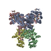 4xz3C 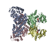 4y8vC 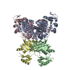 4yajC  4yakC 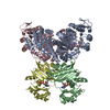 4yb8C 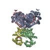 4ybzC 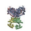 5hbrC C: citing same article ( |
|---|---|
| Similar structure data |


-Links
 PDBj
PDBj


- Assembly
| Deposited unit | 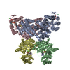
| ||||||||
|---|---|---|---|---|---|---|---|---|---|
| 1 |
| ||||||||
| Unit cell |
|
-Components
-Protein , 2 types, 4 molecules ACBD
| #1: Protein | Mass: 49354.523 Da / Num. of mol.: 2 Source method: isolated from a genetically manipulated source Source: (gene. exp.)  Korarchaeum cryptofilum (strain OPF8) (archaea) Korarchaeum cryptofilum (strain OPF8) (archaea)Gene: Kcr_0198 / Production host:  #2: Protein | Mass: 25676.797 Da / Num. of mol.: 2 Source method: isolated from a genetically manipulated source Source: (gene. exp.) Korarchaeum cryptofilum (strain OPF8) (archaea)Gene: Kcr_0115 / Plasmid: pET17b / Production host: |
|---|
-Non-polymers , 7 types, 457 molecules 


| #3: Chemical | ChemComp-CL /  Mass: 35.453 Da / Num. of mol.: 6 / Source method: obtained synthetically / Formula: Cl Mass: 35.453 Da / Num. of mol.: 6 / Source method: obtained synthetically / Formula: Cl#4: Chemical | ChemComp-HG /  Mass: 200.590 Da / Num. of mol.: 8 / Source method: obtained synthetically / Formula: Hg Mass: 200.590 Da / Num. of mol.: 8 / Source method: obtained synthetically / Formula: Hg#5: Chemical |  Mass: 767.534 Da / Num. of mol.: 2 / Source method: obtained synthetically / Formula: C21H36N7O16P3S Mass: 767.534 Da / Num. of mol.: 2 / Source method: obtained synthetically / Formula: C21H36N7O16P3S#6: Chemical |  Mass: 425.228 Da / Num. of mol.: 2 / Source method: obtained synthetically / Formula: C11H17N5O9P2 Mass: 425.228 Da / Num. of mol.: 2 / Source method: obtained synthetically / Formula: C11H17N5O9P2#7: Chemical |  Mass: 40.078 Da / Num. of mol.: 2 / Source method: obtained synthetically / Formula: Ca Mass: 40.078 Da / Num. of mol.: 2 / Source method: obtained synthetically / Formula: Ca#8: Chemical | ChemComp-NA / |  Mass: 22.990 Da / Num. of mol.: 1 / Source method: obtained synthetically / Formula: Na Mass: 22.990 Da / Num. of mol.: 1 / Source method: obtained synthetically / Formula: Na#9: Water | ChemComp-HOH / | Mass: 18.015 Da / Num. of mol.: 436 / Source method: isolated from a natural source / Formula: H2O |
|---|
-Experimental details
-Experiment
| Experiment | Method: X-RAY DIFFRACTION |
|---|
- Sample preparation
| Crystal | Density Matthews: 2.41 Å3/Da / Density % sol: 48.98 % / Description: rod-like appearance |
|---|---|
| Crystal grow | Temperature: 291 K / Method: vapor diffusion, hanging drop Details: 100 mM Tris/HCl (pH 8.3), 14% (w/v) PEG6000, 30 mM CaCl2, 2 mM HgCl2 |
-Data collection
| Diffraction | Mean temperature: 100 K |
|---|---|
| Diffraction source | Source: SYNCHROTRON / Site: PETRA III, EMBL c/o DESY  / Beamline: P14 (MX2) / Wavelength: 1.23953 Å / Beamline: P14 (MX2) / Wavelength: 1.23953 Å |
| Detector | Type: DECTRIS PILATUS 6M / Detector: PIXEL / Date: Nov 17, 2012 |
| Radiation | Protocol: SINGLE WAVELENGTH / Monochromatic (M) / Laue (L): M / Scattering type: x-ray |
| Radiation wavelength | Wavelength: 1.23953 Å / Relative weight: 1 |
| Reflection | Resolution: 1.9→127.04 Å / Num. all: 112092 / Num. obs: 112092 / % possible obs: 97.8 % / Redundancy: 6.3 % / Rmerge(I) obs: 0.181 / Net I/σ(I): 8.1 |
| Reflection shell | Resolution: 1.9→1.93 Å / Redundancy: 2.7 % / Rmerge(I) obs: 4.278 / Mean I/σ(I) obs: 0.3 / % possible all: 58.1 |
- Processing
| Software |
| |||||||||||||||||||||||||||||||||||||||||||||||||||||||||||||||||||||||||||||||||||||||||||||||||||||||||||||||||||||||||||||||||||||||||||||||||||||||||||||||||||||||||||||||||||||||||||||||||||||||||||||||||||||||||
|---|---|---|---|---|---|---|---|---|---|---|---|---|---|---|---|---|---|---|---|---|---|---|---|---|---|---|---|---|---|---|---|---|---|---|---|---|---|---|---|---|---|---|---|---|---|---|---|---|---|---|---|---|---|---|---|---|---|---|---|---|---|---|---|---|---|---|---|---|---|---|---|---|---|---|---|---|---|---|---|---|---|---|---|---|---|---|---|---|---|---|---|---|---|---|---|---|---|---|---|---|---|---|---|---|---|---|---|---|---|---|---|---|---|---|---|---|---|---|---|---|---|---|---|---|---|---|---|---|---|---|---|---|---|---|---|---|---|---|---|---|---|---|---|---|---|---|---|---|---|---|---|---|---|---|---|---|---|---|---|---|---|---|---|---|---|---|---|---|---|---|---|---|---|---|---|---|---|---|---|---|---|---|---|---|---|---|---|---|---|---|---|---|---|---|---|---|---|---|---|---|---|---|---|---|---|---|---|---|---|---|---|---|---|---|---|---|---|---|
| Refinement | Method to determine structure: MOLECULAR REPLACEMENT / Resolution: 1.9→85.018 Å / SU ML: 0.32 / Cross valid method: FREE R-VALUE / σ(F): 1.33 / Phase error: 27.45 / Stereochemistry target values: ML
| |||||||||||||||||||||||||||||||||||||||||||||||||||||||||||||||||||||||||||||||||||||||||||||||||||||||||||||||||||||||||||||||||||||||||||||||||||||||||||||||||||||||||||||||||||||||||||||||||||||||||||||||||||||||||
| Solvent computation | Shrinkage radii: 0.9 Å / VDW probe radii: 1.11 Å / Solvent model: FLAT BULK SOLVENT MODEL | |||||||||||||||||||||||||||||||||||||||||||||||||||||||||||||||||||||||||||||||||||||||||||||||||||||||||||||||||||||||||||||||||||||||||||||||||||||||||||||||||||||||||||||||||||||||||||||||||||||||||||||||||||||||||
| Refinement step | Cycle: LAST / Resolution: 1.9→85.018 Å
| |||||||||||||||||||||||||||||||||||||||||||||||||||||||||||||||||||||||||||||||||||||||||||||||||||||||||||||||||||||||||||||||||||||||||||||||||||||||||||||||||||||||||||||||||||||||||||||||||||||||||||||||||||||||||
| Refine LS restraints |
| |||||||||||||||||||||||||||||||||||||||||||||||||||||||||||||||||||||||||||||||||||||||||||||||||||||||||||||||||||||||||||||||||||||||||||||||||||||||||||||||||||||||||||||||||||||||||||||||||||||||||||||||||||||||||
| LS refinement shell |
|
