 Movie
Movie Controller

[English] 日本語
 Yorodumi
- PDB-4xoq: F420 complex of coenzyme F420:L-glutamate ligase (FbiB) from Myco... -
+ Open data

- Basic information
| Entry | Database: PDB / ID: 4xoq | ||||||
|---|---|---|---|---|---|---|---|
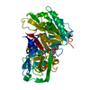
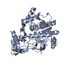
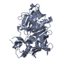
| Title | F420 complex of coenzyme F420:L-glutamate ligase (FbiB) from Mycobacterium tuberculosis (C-terminal domain) | ||||||
 Components Components | Coenzyme F420:L-glutamate ligase | ||||||
 Keywords Keywords | UNKNOWN FUNCTION / NADH-oxidase fold / FMN- and F420-binding | ||||||
| Function / homology |  Function and homology information Function and homology informationdehydro coenzyme F420 reductase / oxidoreductase activity, acting on the CH-CH group of donors, with a flavin as acceptor / coenzyme gamma-F420-2 biosynthetic process / coenzyme F420-0:L-glutamate ligase / coenzyme F420-1:gamma-L-glutamate ligase / coenzyme F420-0:L-glutamate ligase activity / coenzyme F420-1:gamma-L-glutamate ligase activity / F420-0 metabolic process / GTP binding / metal ion binding / plasma membrane Similarity search - Function | ||||||
| Biological species |   Mycobacterium tuberculosis (bacteria) Mycobacterium tuberculosis (bacteria) | ||||||
| Method |  X-RAY DIFFRACTION / SYNCHROTRON / MOLECULAR REPLACEMENT / Resolution: 2.05 Å X-RAY DIFFRACTION / SYNCHROTRON / MOLECULAR REPLACEMENT / Resolution: 2.05 Å | ||||||
 Authors Authors | Rehan, A.M. / Bashiri, G. / Baker, H.M. / Baker, E.N. / Squire, C.J. | ||||||
 Citation Citation | Journal: J.Biol.Chem. / Year: 2016 Title: Elongation of the Poly-gamma-glutamate Tail of F420 Requires Both Domains of the F420: gamma-Glutamyl Ligase (FbiB) of Mycobacterium tuberculosis. Authors: Bashiri, G. / Rehan, A.M. / Sreebhavan, S. / Baker, H.M. / Baker, E.N. / Squire, C.J. | ||||||
| History |
|
- Structure visualization
| Structure viewer | Molecule: MolmilJmol/JSmol |
|---|
- Downloads & links
-Download
| PDBx/mmCIF format | 4xoq.cif.gz | 316.6 KB | Display | PDBx/mmCIF format |
|---|---|---|---|---|
| PDB format | pdb4xoq.ent.gz | 256.2 KB | Display | PDB format |
| PDBx/mmJSON format | 4xoq.json.gz | Tree view | PDBx/mmJSON format | |
| Others |  Other downloads Other downloads |
-Validation report
| Arichive directory | https://data.pdbj.org/pub/pdb/validation_reports/xo/4xoqftp://data.pdbj.org/pub/pdb/validation_reports/xo/4xoq | HTTPS FTP |
|---|
-Related structure data
| Related structure data | 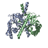 4xomSC  4xooC S: Starting model for refinement C: citing same article ( |
|---|---|
| Similar structure data |


-Links
 PDBj
PDBj- Assembly
| Deposited unit | 
| ||||||||
|---|---|---|---|---|---|---|---|---|---|
| 1 | 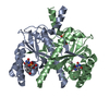
| ||||||||
| 2 | 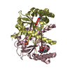
| ||||||||
| Unit cell |
| ||||||||
| Components on special symmetry positions |
|
-Components
| #1: Protein | Mass: 22352.562 Da / Num. of mol.: 4 / Fragment: UNP residues 245-448 Source method: isolated from a genetically manipulated source Source: (gene. exp.) Mycobacterium tuberculosis (strain ATCC 25618 / H37Rv) (bacteria)Strain: ATCC 25618 / H37Rv / Gene: fbiB, Rv3262 / Production host: References: UniProt: P9WP79, coenzyme F420-0:L-glutamate ligase, coenzyme F420-1:gamma-L-glutamate ligase #2: Chemical | ChemComp-F42 /   Mass: 773.593 Da / Num. of mol.: 4 / Source method: obtained synthetically / Formula: C29H36N5O18P Mass: 773.593 Da / Num. of mol.: 4 / Source method: obtained synthetically / Formula: C29H36N5O18P#3: Chemical |   Mass: 96.063 Da / Num. of mol.: 3 / Source method: obtained synthetically / Formula: SO4 Mass: 96.063 Da / Num. of mol.: 3 / Source method: obtained synthetically / Formula: SO4#4: Water | ChemComp-HOH / |  Mass: 18.015 Da / Num. of mol.: 398 / Source method: isolated from a natural source / Formula: H2O Mass: 18.015 Da / Num. of mol.: 398 / Source method: isolated from a natural source / Formula: H2O |
|---|
-Experimental details
-Experiment
| Experiment | Method: X-RAY DIFFRACTION |
|---|
- Sample preparation
| Crystal | Density Matthews: 2.65 Å3/Da / Density % sol: 53.67 % |
|---|---|
| Crystal grow | Temperature: 291 K / Method: vapor diffusion, hanging drop / Details: PEG 3350, lithium sulfate |
-Data collection
| Diffraction | Mean temperature: 100 K |
|---|---|
| Diffraction source | Source: SYNCHROTRON / Site: Australian Synchrotron  / Beamline: MX2 / Wavelength: 0.9537 Å / Beamline: MX2 / Wavelength: 0.9537 Å |
| Detector | Type: ADSC QUANTUM 315r / Detector: CCD / Date: Nov 26, 2012 |
| Radiation | Protocol: SINGLE WAVELENGTH / Monochromatic (M) / Laue (L): M / Scattering type: x-ray |
| Radiation wavelength | Wavelength: 0.9537 Å / Relative weight: 1 |
| Reflection | Resolution: 2.05→48.3 Å / Num. obs: 60778 / % possible obs: 100 % / Redundancy: 14.6 % / CC1/2: 0.998 / Net I/σ(I): 18.5 |
| Reflection shell | Resolution: 2.05→2.1 Å / Redundancy: 14.1 % / Mean I/σ(I) obs: 2 / CC1/2: 0.659 / % possible all: 99.8 |
- Processing
| Software |
| ||||||||||||||||||||||||||||||||||||||||||||||||||||||||||||||||||||||||||||||||||||||||||||||||||||||||||||||||||||||||||||||||||||||||||||||||||||||||||||||||||||||||||||||||||||||
|---|---|---|---|---|---|---|---|---|---|---|---|---|---|---|---|---|---|---|---|---|---|---|---|---|---|---|---|---|---|---|---|---|---|---|---|---|---|---|---|---|---|---|---|---|---|---|---|---|---|---|---|---|---|---|---|---|---|---|---|---|---|---|---|---|---|---|---|---|---|---|---|---|---|---|---|---|---|---|---|---|---|---|---|---|---|---|---|---|---|---|---|---|---|---|---|---|---|---|---|---|---|---|---|---|---|---|---|---|---|---|---|---|---|---|---|---|---|---|---|---|---|---|---|---|---|---|---|---|---|---|---|---|---|---|---|---|---|---|---|---|---|---|---|---|---|---|---|---|---|---|---|---|---|---|---|---|---|---|---|---|---|---|---|---|---|---|---|---|---|---|---|---|---|---|---|---|---|---|---|---|---|---|---|
| Refinement | Method to determine structure: MOLECULAR REPLACEMENT Starting model: 4XOM Resolution: 2.05→48.3 Å / Cor.coef. Fo:Fc: 0.943 / Cor.coef. Fo:Fc free: 0.931 / SU B: 10.923 / SU ML: 0.146 / Cross valid method: THROUGHOUT / ESU R: 0.198 / ESU R Free: 0.171 / Stereochemistry target values: MAXIMUM LIKELIHOOD / Details: HYDROGENS HAVE BEEN ADDED IN THE RIDING POSITIONS
| ||||||||||||||||||||||||||||||||||||||||||||||||||||||||||||||||||||||||||||||||||||||||||||||||||||||||||||||||||||||||||||||||||||||||||||||||||||||||||||||||||||||||||||||||||||||
| Solvent computation | Ion probe radii: 0.8 Å / Shrinkage radii: 0.8 Å / VDW probe radii: 1.2 Å / Solvent model: MASK | ||||||||||||||||||||||||||||||||||||||||||||||||||||||||||||||||||||||||||||||||||||||||||||||||||||||||||||||||||||||||||||||||||||||||||||||||||||||||||||||||||||||||||||||||||||||
| Displacement parameters | Biso mean: 32.848 Å2
| ||||||||||||||||||||||||||||||||||||||||||||||||||||||||||||||||||||||||||||||||||||||||||||||||||||||||||||||||||||||||||||||||||||||||||||||||||||||||||||||||||||||||||||||||||||||
| Refinement step | Cycle: 1 / Resolution: 2.05→48.3 Å
| ||||||||||||||||||||||||||||||||||||||||||||||||||||||||||||||||||||||||||||||||||||||||||||||||||||||||||||||||||||||||||||||||||||||||||||||||||||||||||||||||||||||||||||||||||||||
| Refine LS restraints |
|