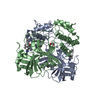
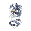
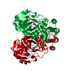
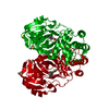
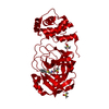
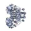
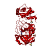
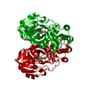
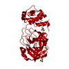
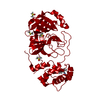
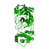
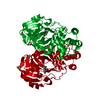
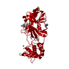
Entry Database : PDB / ID : 4xfqTitle Crystal Structure Basis for PEDV 3C Like Protease PEDV main protease Keywords / / Function / homology Function Domain/homology Component
/ / / / / / / / / / / / / / / / / / / / / / / / / / / / / / / / / / / / / / / / / / / / / / / / / / / / / / / / / / / / / / / / / / / / / / / / / / / / / / / / / / / / / / / / / / / / / / / / / / / / / / / / / / / / / / / / / / / / / / / / / / / / / / / / / / / / / / / / / / / / / Biological species Method / / / Resolution : 1.65 Å Authors Ye, G. / Fu, Z.F. / Peng, G.Q. Journal : Virology / Year : 2016Title : Structural basis for the dimerization and substrate recognition specificity of porcine epidemic diarrhea virus 3C-like protease.Authors : Ye, G. / Deng, F. / Shen, Z. / Luo, R. / Zhao, L. / Xiao, S. / Fu, Z.F. / Peng, G. History Deposition Dec 28, 2014 Deposition site / Processing site Revision 1.0 Jan 20, 2016 Provider / Type Revision 1.1 Jun 22, 2016 Group Revision 1.2 Nov 8, 2023 Group Data collection / Database references ... Data collection / Database references / Derived calculations / Refinement description Category chem_comp_atom / chem_comp_bond ... chem_comp_atom / chem_comp_bond / citation / database_2 / pdbx_initial_refinement_model / pdbx_struct_oper_list / refine / software Item _citation.journal_id_CSD / _database_2.pdbx_DOI ... _citation.journal_id_CSD / _database_2.pdbx_DOI / _database_2.pdbx_database_accession / _pdbx_struct_oper_list.symmetry_operation / _refine.pdbx_method_to_determine_struct / _software.name
Show all Show less
 Movie
Movie Controller


 Open data
 Basic information
 Components
 Keywords
 Function and homology information
 Porcine epidemic diarrhea virus
 X-RAY DIFFRACTION /
  Authors
 Citation
 Structure visualization
 Downloads & links
 Other downloads
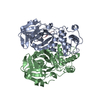


 PDBj
PDBj


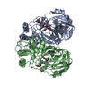
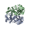
 Assembly

 Mass: 18.015 Da / Num. of mol.: 461 / Source method: isolated from a natural source / Formula: H2O
 Sample preparation
 / Beamline: BL17U / Wavelength: 0.97917 Å
 Processing