 Movie
Movie Controller

[English] 日本語
 Yorodumi
- PDB-4wsp: Racemic crystal structure of Rv1738 from Mycobacterium tuberculos... -
+ Open data

- Basic information
| Entry | Database: PDB / ID: 4wsp | ||||||
|---|---|---|---|---|---|---|---|
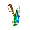
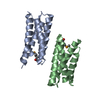
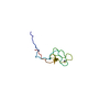
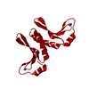
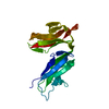
| Title | Racemic crystal structure of Rv1738 from Mycobacterium tuberculosis (Form-I) | ||||||
 Components Components | protein DL-Rv1738 | ||||||
 Keywords Keywords | DE NOVO PROTEIN / hypoxic response | ||||||
| Function / homology |  Function and homology information Function and homology information | ||||||
| Biological species |   Mycobacterium tuberculosis (bacteria) Mycobacterium tuberculosis (bacteria) | ||||||
| Method |  X-RAY DIFFRACTION / SYNCHROTRON / MOLECULAR REPLACEMENT / Resolution: 1.65 Å X-RAY DIFFRACTION / SYNCHROTRON / MOLECULAR REPLACEMENT / Resolution: 1.65 Å | ||||||
 Authors Authors | Bunker, R.D. / Mandal, K. / Kent, S.B.H. / Baker, E.N. | ||||||
 Citation Citation | Journal: Proc.Natl.Acad.Sci.USA / Year: 2015 Title: A functional role of Rv1738 in Mycobacterium tuberculosis persistence suggested by racemic protein crystallography. Authors: Bunker, R.D. / Mandal, K. / Bashiri, G. / Chaston, J.J. / Pentelute, B.L. / Lott, J.S. / Kent, S.B. / Baker, E.N. | ||||||
| History |
|
- Structure visualization
| Structure viewer | Molecule: MolmilJmol/JSmol |
|---|
- Downloads & links
-Download
| PDBx/mmCIF format | 4wsp.cif.gz | 60.4 KB | Display | PDBx/mmCIF format |
|---|---|---|---|---|
| PDB format | pdb4wsp.ent.gz | 44.4 KB | Display | PDB format |
| PDBx/mmJSON format | 4wsp.json.gz | Tree view | PDBx/mmJSON format | |
| Others |  Other downloads Other downloads |
-Validation report
| Summary document | 4wsp_validation.pdf.gz | 422 KB | Display | wwPDB validaton report |
|---|---|---|---|---|
| Full document | 4wsp_full_validation.pdf.gz | 423.5 KB | Display | |
| Data in XML | 4wsp_validation.xml.gz | 4.8 KB | Display | |
| Data in CIF | 4wsp_validation.cif.gz | 6 KB | Display | |
| Arichive directory | https://data.pdbj.org/pub/pdb/validation_reports/ws/4wspftp://data.pdbj.org/pub/pdb/validation_reports/ws/4wsp | HTTPS FTP |
-Related structure data
| Related structure data | 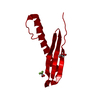 4wpySC S: Starting model for refinement C: citing same article ( |
|---|---|
| Similar structure data |

-Links
 PDBj
PDBj
- Assembly
| Deposited unit | 
| ||||||||
|---|---|---|---|---|---|---|---|---|---|
| 1 |
| ||||||||
| Unit cell |
| ||||||||
| Details | biological unit is the same as asym. |
-Components
| #1: Protein | Mass: 10624.082 Da / Num. of mol.: 1 / Source method: obtained synthetically / Source: (synth.) Mycobacterium tuberculosis (bacteria) / References: UniProt: P9WLS3 |
|---|---|
| #2: Chemical | ChemComp-CL /   Mass: 35.453 Da / Num. of mol.: 1 / Source method: obtained synthetically / Formula: Cl Mass: 35.453 Da / Num. of mol.: 1 / Source method: obtained synthetically / Formula: Cl |
| #3: Water | ChemComp-HOH /  Mass: 18.015 Da / Num. of mol.: 18 / Source method: isolated from a natural source / Formula: H2O Mass: 18.015 Da / Num. of mol.: 18 / Source method: isolated from a natural source / Formula: H2O |
-Experimental details
-Experiment
| Experiment | Method: X-RAY DIFFRACTION / Number of used crystals: 1 |
|---|
- Sample preparation
| Crystal | Density Matthews: 1.66 Å3/Da / Density % sol: 26 % |
|---|---|
| Crystal grow | Temperature: 292 K / Method: vapor diffusion, hanging drop / pH: 5.5 Details: 1 microlitre of protein solution containing 9 mg/ml L-Rv1738 and 9 mg/ml D-Rv1738 in water was mixed with 1 microlitre of well solution containing 0.1 M BIS-TRIS pH 5.5, 0.2 M NaCl, 25 % (w/v) PEG 3350 |
-Data collection
| Diffraction | Mean temperature: 100 K | |||||||||||||||||||||||||||
|---|---|---|---|---|---|---|---|---|---|---|---|---|---|---|---|---|---|---|---|---|---|---|---|---|---|---|---|---|
| Diffraction source | Source: SYNCHROTRON / Site: APS  / Beamline: 23-BM-B / Wavelength: 0.97934 Å / Beamline: 23-BM-B / Wavelength: 0.97934 Å | |||||||||||||||||||||||||||
| Detector | Type: MARMOSAIC 300 mm CCD / Detector: CCD / Date: Apr 23, 2008 | |||||||||||||||||||||||||||
| Radiation | Monochromator: Si(111) / Protocol: SINGLE WAVELENGTH / Monochromatic (M) / Laue (L): M / Scattering type: x-ray | |||||||||||||||||||||||||||
| Radiation wavelength | Wavelength: 0.97934 Å / Relative weight: 1 | |||||||||||||||||||||||||||
| Reflection | Resolution: 1.65→47.65 Å / Num. obs: 17584 / % possible obs: 98.1 % / Redundancy: 2.8 % / Biso Wilson estimate: 49.07 Å2 / CC1/2: 0.998 / Rmerge(I) obs: 0.057 / Rpim(I) all: 0.041 / Net I/σ(I): 9.4 / Num. measured all: 49522 | |||||||||||||||||||||||||||
| Reflection shell | Diffraction-ID: 1 / Rejects: _
|
- Processing
| Software |
| ||||||||||||||||||||||||||||||||||||||||||||||||||||||||||||||||||||||
|---|---|---|---|---|---|---|---|---|---|---|---|---|---|---|---|---|---|---|---|---|---|---|---|---|---|---|---|---|---|---|---|---|---|---|---|---|---|---|---|---|---|---|---|---|---|---|---|---|---|---|---|---|---|---|---|---|---|---|---|---|---|---|---|---|---|---|---|---|---|---|---|
| Refinement | Method to determine structure: MOLECULAR REPLACEMENT Starting model: 4WPY Resolution: 1.65→39.344 Å / SU ML: 0.2 / Cross valid method: THROUGHOUT / σ(F): 1.34 / Phase error: 23.53 / Stereochemistry target values: ML
| ||||||||||||||||||||||||||||||||||||||||||||||||||||||||||||||||||||||
| Solvent computation | Shrinkage radii: 0.8 Å / VDW probe radii: 1.1 Å / Solvent model: FLAT BULK SOLVENT MODEL | ||||||||||||||||||||||||||||||||||||||||||||||||||||||||||||||||||||||
| Displacement parameters | Biso max: 137.13 Å2 / Biso mean: 49.4724 Å2 / Biso min: 22.03 Å2 | ||||||||||||||||||||||||||||||||||||||||||||||||||||||||||||||||||||||
| Refinement step | Cycle: final / Resolution: 1.65→39.344 Å
| ||||||||||||||||||||||||||||||||||||||||||||||||||||||||||||||||||||||
| Refine LS restraints |
| ||||||||||||||||||||||||||||||||||||||||||||||||||||||||||||||||||||||
| LS refinement shell | Refine-ID: X-RAY DIFFRACTION / Total num. of bins used: 9
|
