 Movie
Movie Controller

[English] 日本語
 Yorodumi
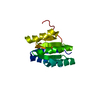
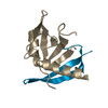
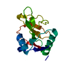
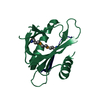
- PDB-4w8h: Crystal structure of the TIR domain of the Toll-related Receptor ... -
+ Open data

- Basic information
| Entry | Database: PDB / ID: 4w8h | ||||||
|---|---|---|---|---|---|---|---|
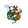
| Title | Crystal structure of the TIR domain of the Toll-related Receptor TRR-2 from the lower metazoan Hydra magnipapillata (crystal form II) | ||||||
 Components Components |
| ||||||
 Keywords Keywords | SIGNALING PROTEIN / Flavodoxin-like / Toll/Interleukin receptor TIR domain | ||||||
| Function / homology |  Function and homology information Function and homology information | ||||||
| Biological species |  synthetic construct (others) | ||||||
| Method |  X-RAY DIFFRACTION / SYNCHROTRON / MOLECULAR REPLACEMENT / Resolution: 1.14 Å X-RAY DIFFRACTION / SYNCHROTRON / MOLECULAR REPLACEMENT / Resolution: 1.14 Å | ||||||
 Authors Authors | Weisse, R.H.-J. / Scheidig, A.J. | ||||||
 Citation Citation | Journal: To Be Published Title: Crystal structure of the TIR domain of the Toll-related Receptor TRR-2 from the lower metazoan Hydra magnipapillata (crystal form II) Authors: Weisse, R.H.-J. / Scheidig, A.J. | ||||||
| History |
|
- Structure visualization
| Structure viewer | Molecule: MolmilJmol/JSmol |
|---|
- Downloads & links
-Download
| PDBx/mmCIF format | 4w8h.cif.gz | 101.9 KB | Display | PDBx/mmCIF format |
|---|---|---|---|---|
| PDB format | pdb4w8h.ent.gz | 79.1 KB | Display | PDB format |
| PDBx/mmJSON format | 4w8h.json.gz | Tree view | PDBx/mmJSON format | |
| Others |  Other downloads Other downloads |
-Validation report
| Arichive directory | https://data.pdbj.org/pub/pdb/validation_reports/w8/4w8hftp://data.pdbj.org/pub/pdb/validation_reports/w8/4w8h | HTTPS FTP |
|---|
-Related structure data
| Related structure data |  4w8gS S: Starting model for refinement |
|---|---|
| Similar structure data |


-Links
 PDBj
PDBj
- Assembly
| Deposited unit | 
| ||||||||
|---|---|---|---|---|---|---|---|---|---|
| 1 |
| ||||||||
| Unit cell |
|
-Components
| #1: Protein | Mass: 15330.566 Da / Num. of mol.: 1 / Fragment: UNP residues 95-221 Source method: isolated from a genetically manipulated source Source: (gene. exp.)  |
|---|---|
| #2: Protein/peptide | Mass: 846.896 Da / Num. of mol.: 1 Source method: isolated from a genetically manipulated source Source: (gene. exp.) synthetic construct (others) / Plasmid: pET19b / Production host: |
| #3: Chemical | ChemComp-CL /   Mass: 35.453 Da / Num. of mol.: 1 / Source method: obtained synthetically / Formula: Cl Mass: 35.453 Da / Num. of mol.: 1 / Source method: obtained synthetically / Formula: Cl |
| #4: Water | ChemComp-HOH /  Mass: 18.015 Da / Num. of mol.: 162 / Source method: isolated from a natural source / Formula: H2O Mass: 18.015 Da / Num. of mol.: 162 / Source method: isolated from a natural source / Formula: H2O |
-Experimental details
-Experiment
| Experiment | Method: X-RAY DIFFRACTION |
|---|
- Sample preparation
| Crystal | Density Matthews: 1.87 Å3/Da / Density % sol: 34.35 % / Description: short, needle-like appearance |
|---|---|
| Crystal grow | Temperature: 293 K / Method: vapor diffusion, hanging drop Details: 100mM NaOAc (pH 5;8), 350mM MgCl2, 18% (w/v) PEG 3350 |
-Data collection
| Diffraction | Mean temperature: 100 K |
|---|---|
| Diffraction source | Source: SYNCHROTRON / Site: BESSY  / Beamline: 14.1 / Wavelength: 0.918069 Å / Beamline: 14.1 / Wavelength: 0.918069 Å |
| Detector | Type: DECTRIS PILATUS 6M / Detector: PIXEL / Date: Apr 18, 2013 |
| Radiation | Protocol: SINGLE WAVELENGTH / Monochromatic (M) / Laue (L): M / Scattering type: x-ray |
| Radiation wavelength | Wavelength: 0.918069 Å / Relative weight: 1 |
| Reflection | Resolution: 1.14→31.79 Å / Num. all: 42226 / Num. obs: 42226 / % possible obs: 97.5 % / Redundancy: 5.8 % / Rmerge(I) obs: 0.112 / Net I/σ(I): 9.1 |
| Reflection shell | Resolution: 1.14→1.16 Å / Redundancy: 3.6 % / Rmerge(I) obs: 1.659 / Mean I/σ(I) obs: 0.7 / % possible all: 96.1 |
- Processing
| Software |
| ||||||||||||||||||||||||||||||||||||||||||||||||||||||||||||||||||||||||||||||||||||||||||||||||||||||||||||||||
|---|---|---|---|---|---|---|---|---|---|---|---|---|---|---|---|---|---|---|---|---|---|---|---|---|---|---|---|---|---|---|---|---|---|---|---|---|---|---|---|---|---|---|---|---|---|---|---|---|---|---|---|---|---|---|---|---|---|---|---|---|---|---|---|---|---|---|---|---|---|---|---|---|---|---|---|---|---|---|---|---|---|---|---|---|---|---|---|---|---|---|---|---|---|---|---|---|---|---|---|---|---|---|---|---|---|---|---|---|---|---|---|---|---|
| Refinement | Method to determine structure: MOLECULAR REPLACEMENT Starting model: 4W8G Resolution: 1.14→31.785 Å / SU ML: 0.15 / Cross valid method: THROUGHOUT / σ(F): 1.36 / Phase error: 20.14 / Stereochemistry target values: ML
| ||||||||||||||||||||||||||||||||||||||||||||||||||||||||||||||||||||||||||||||||||||||||||||||||||||||||||||||||
| Solvent computation | Shrinkage radii: 0.9 Å / VDW probe radii: 1.11 Å / Solvent model: FLAT BULK SOLVENT MODEL | ||||||||||||||||||||||||||||||||||||||||||||||||||||||||||||||||||||||||||||||||||||||||||||||||||||||||||||||||
| Refinement step | Cycle: LAST / Resolution: 1.14→31.785 Å
| ||||||||||||||||||||||||||||||||||||||||||||||||||||||||||||||||||||||||||||||||||||||||||||||||||||||||||||||||
| Refine LS restraints |
| ||||||||||||||||||||||||||||||||||||||||||||||||||||||||||||||||||||||||||||||||||||||||||||||||||||||||||||||||
| LS refinement shell |
|
