 Movie
Movie Controller

[English] 日本語
 Yorodumi
- PDB-4v1z: The 3-D structure of the cellobiohydrolase, Cel7A, from Aspergill... -
+ Open data

- Basic information
| Entry | Database: PDB / ID: 4v1z | |||||||||
|---|---|---|---|---|---|---|---|---|---|---|
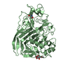
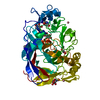
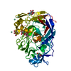
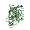
| Title | The 3-D structure of the cellobiohydrolase, Cel7A, from Aspergillus fumigatus | |||||||||
 Components Components | CELLOBIOHYDROLASE | |||||||||
 Keywords Keywords | HYDROLASE / CELLULASE / BIOFUELS / CARBOHYDRATE-ACTIVE ENZYME / THERMAL 2 STABILITY | |||||||||
| Function / homology |  Function and homology information Function and homology informationcellulose 1,4-beta-cellobiosidase (non-reducing end) / cellulose 1,4-beta-cellobiosidase activity / glucan catabolic process / cellulose binding / cellulose catabolic process / extracellular region Similarity search - Function | |||||||||
| Biological species |  | |||||||||
| Method |  X-RAY DIFFRACTION / SYNCHROTRON / MOLECULAR REPLACEMENT / Resolution: 1.78 Å X-RAY DIFFRACTION / SYNCHROTRON / MOLECULAR REPLACEMENT / Resolution: 1.78 Å | |||||||||
 Authors Authors | Moroz, O.V. / Maranta, M. / Shaghasi, T. / Harris, P.V. / Wilson, K.S. / Davies, G.J. | |||||||||
 Citation Citation | Journal: Acta Crystallogr.,Sect.F / Year: 2015 Title: The Three-Dimensional Structure of the Cellobiohydrolase Cel7A from Aspergillus Fumigatus at 1.5 A Resolution Authors: Moroz, O.V. / Maranta, M. / Shaghasi, T. / Harris, P.V. / Wilson, K.S. / Davies, G.J. | |||||||||
| History |
|
- Structure visualization
| Structure viewer | Molecule: MolmilJmol/JSmol |
|---|
- Downloads & links
-Download
| PDBx/mmCIF format | 4v1z.cif.gz | 110.7 KB | Display | PDBx/mmCIF format |
|---|---|---|---|---|
| PDB format | pdb4v1z.ent.gz | 84.1 KB | Display | PDB format |
| PDBx/mmJSON format | 4v1z.json.gz | Tree view | PDBx/mmJSON format | |
| Others |  Other downloads Other downloads |
-Validation report
| Arichive directory | https://data.pdbj.org/pub/pdb/validation_reports/v1/4v1zftp://data.pdbj.org/pub/pdb/validation_reports/v1/4v1z | HTTPS FTP |
|---|
-Related structure data
| Related structure data | 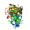 4v20C  1q9hS C: citing same article ( S: Starting model for refinement |
|---|---|
| Similar structure data |


-Links
 PDBj
PDBj- Assembly
| Deposited unit | 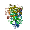
| ||||||||
|---|---|---|---|---|---|---|---|---|---|
| 1 |
| ||||||||
| Unit cell |
|
-Components
| #1: Protein | Mass: 47137.566 Da / Num. of mol.: 1 / Fragment: UNP RESIDUES 27-466 Source method: isolated from a genetically manipulated source Source: (gene. exp.) References: UniProt: Q4WM08, cellulose 1,4-beta-cellobiosidase (non-reducing end) | ||||
|---|---|---|---|---|---|
| #2: Sugar | ChemComp-NAG /   Type: D-saccharide, beta linking / Mass: 221.208 Da / Num. of mol.: 1 Type: D-saccharide, beta linking / Mass: 221.208 Da / Num. of mol.: 1Source method: isolated from a genetically manipulated source Formula: C8H15NO6 | ||||
| #3: Chemical |   Mass: 65.409 Da / Num. of mol.: 2 / Source method: obtained synthetically / Formula: Zn Mass: 65.409 Da / Num. of mol.: 2 / Source method: obtained synthetically / Formula: Zn#4: Water | ChemComp-HOH / |  Mass: 18.015 Da / Num. of mol.: 470 / Source method: isolated from a natural source / Formula: H2O Mass: 18.015 Da / Num. of mol.: 470 / Source method: isolated from a natural source / Formula: H2OHas protein modification | Y | |
-Experimental details
-Experiment
| Experiment | Method: X-RAY DIFFRACTION |
|---|
- Sample preparation
| Crystal | Density Matthews: 2.54 Å3/Da / Density % sol: 51 % / Description: NONE |
|---|---|
| Crystal grow | Details: PACT B12 20% PEG6K, 10 MM ZNCL2, MES PH 6.0, OPTIMIZED MANUALLY |
-Data collection
| Diffraction | Mean temperature: 120 K |
|---|---|
| Diffraction source | Source: SYNCHROTRON / Site: Diamond  / Beamline: I03 / Type: DIAMOND / Wavelength: 0.98 / Beamline: I03 / Type: DIAMOND / Wavelength: 0.98 |
| Detector | Date: Jul 11, 2011 |
| Radiation | Protocol: SINGLE WAVELENGTH / Monochromatic (M) / Laue (L): M / Scattering type: x-ray |
| Radiation wavelength | Wavelength: 0.98 Å / Relative weight: 1 |
| Reflection | Resolution: 1.78→65.69 Å / Num. obs: 44036 / % possible obs: 99.3 % / Observed criterion σ(I): 2 / Redundancy: 5.9 % / Rmerge(I) obs: 0.1 / Net I/σ(I): 10.7 |
| Reflection shell | Resolution: 1.78→1.88 Å / Redundancy: 6.1 % / Rmerge(I) obs: 0.68 / Mean I/σ(I) obs: 2.9 / % possible all: 98.2 |
- Processing
| Software |
| ||||||||||||||||||||||||||||||||||||||||||||||||||||||||||||||||||||||||||||||||||||||||||||||||||||||||||||||||||||||||||||||||||||||||||||||||||||||||||||||||||||||||||||||||||||||
|---|---|---|---|---|---|---|---|---|---|---|---|---|---|---|---|---|---|---|---|---|---|---|---|---|---|---|---|---|---|---|---|---|---|---|---|---|---|---|---|---|---|---|---|---|---|---|---|---|---|---|---|---|---|---|---|---|---|---|---|---|---|---|---|---|---|---|---|---|---|---|---|---|---|---|---|---|---|---|---|---|---|---|---|---|---|---|---|---|---|---|---|---|---|---|---|---|---|---|---|---|---|---|---|---|---|---|---|---|---|---|---|---|---|---|---|---|---|---|---|---|---|---|---|---|---|---|---|---|---|---|---|---|---|---|---|---|---|---|---|---|---|---|---|---|---|---|---|---|---|---|---|---|---|---|---|---|---|---|---|---|---|---|---|---|---|---|---|---|---|---|---|---|---|---|---|---|---|---|---|---|---|---|---|
| Refinement | Method to determine structure: MOLECULAR REPLACEMENT Starting model: PDB ENTRY 1Q9H Resolution: 1.78→67.85 Å / Cor.coef. Fo:Fc: 0.973 / Cor.coef. Fo:Fc free: 0.957 / SU B: 2.485 / SU ML: 0.074 / Cross valid method: THROUGHOUT / ESU R: 0.097 / ESU R Free: 0.102 / Stereochemistry target values: MAXIMUM LIKELIHOOD / Details: HYDROGENS HAVE BEEN ADDED IN THE RIDING POSITIONS.
| ||||||||||||||||||||||||||||||||||||||||||||||||||||||||||||||||||||||||||||||||||||||||||||||||||||||||||||||||||||||||||||||||||||||||||||||||||||||||||||||||||||||||||||||||||||||
| Solvent computation | Ion probe radii: 0.8 Å / Shrinkage radii: 0.8 Å / VDW probe radii: 1.2 Å / Solvent model: MASK | ||||||||||||||||||||||||||||||||||||||||||||||||||||||||||||||||||||||||||||||||||||||||||||||||||||||||||||||||||||||||||||||||||||||||||||||||||||||||||||||||||||||||||||||||||||||
| Displacement parameters | Biso mean: 21.699 Å2
| ||||||||||||||||||||||||||||||||||||||||||||||||||||||||||||||||||||||||||||||||||||||||||||||||||||||||||||||||||||||||||||||||||||||||||||||||||||||||||||||||||||||||||||||||||||||
| Refinement step | Cycle: LAST / Resolution: 1.78→67.85 Å
| ||||||||||||||||||||||||||||||||||||||||||||||||||||||||||||||||||||||||||||||||||||||||||||||||||||||||||||||||||||||||||||||||||||||||||||||||||||||||||||||||||||||||||||||||||||||
| Refine LS restraints |
|