 Movie
Movie Controller

[English] 日本語
 Yorodumi
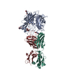
- PDB-4uw8: Structure of the carboxy-terminal domain of the bacteriophage T5 ... -
+ Open data

- Basic information
| Entry | Database: PDB / ID: 4uw8 | ||||||
|---|---|---|---|---|---|---|---|
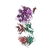
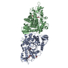
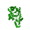
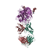
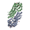
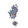
| Title | Structure of the carboxy-terminal domain of the bacteriophage T5 L- shaped tail fiber with its intra-molecular chaperone domain | ||||||
 Components Components | L-SHAPED TAIL FIBER PROTEIN | ||||||
 Keywords Keywords | VIRAL PROTEIN / BACTERIAL VIRUSES / CAUDOVIRALES / SIPHOVIRIDAE / INFECTION | ||||||
| Function / homology |  Function and homology information Function and homology informationlipopolysaccharide-mediated virion attachment to host cell / virus tail, fiber / adhesion receptor-mediated virion attachment to host cell / Hydrolases; Acting on peptide bonds (peptidases); Serine endopeptidases / serine-type peptidase activity / symbiont entry into host cell / proteolysis Similarity search - Function | ||||||
| Biological species |  ENTEROBACTERIA PHAGE T5 (virus) ENTEROBACTERIA PHAGE T5 (virus) | ||||||
| Method |  X-RAY DIFFRACTION / SYNCHROTRON / MOLECULAR REPLACEMENT / Resolution: 2.52 Å X-RAY DIFFRACTION / SYNCHROTRON / MOLECULAR REPLACEMENT / Resolution: 2.52 Å | ||||||
 Authors Authors | Garcia-Doval, C. / Luque, D. / Caston, J.R. / Otero, J.M. / Llamas-Saiz, A.L. / Boulanger, P. / van Raaij, M.J. | ||||||
 Citation Citation | Journal: Viruses / Year: 2015 Title: Structure of the Receptor-Binding Carboxy-Terminal Domain of the Bacteriophage T5 L-Shaped Tail Fibre with and without Its Intra-Molecular Chaperone. Authors: Garcia-Doval, C. / Caston, J.R. / Luque, D. / Granell, M. / Otero, J.M. / Llamas-Saiz, A.L. / Renouard, M. / Boulanger, P. / van Raaij, M.J. #1: Journal: Acta Crystallogr.,Sect.F / Year: 2013 Title: Crystallization of the C-Terminal Domain of the Bacteriophage T5 L-Shaped Fibre. Authors: Garcia-Doval, C. / Luque, D. / Caston, J.R. / Boulanger, P. / van Raaij, M.J. | ||||||
| History |
|
- Structure visualization
| Structure viewer | Molecule: MolmilJmol/JSmol |
|---|
- Downloads & links
-Download
| PDBx/mmCIF format | 4uw8.cif.gz | 693.4 KB | Display | PDBx/mmCIF format |
|---|---|---|---|---|
| PDB format | pdb4uw8.ent.gz | 575.6 KB | Display | PDB format |
| PDBx/mmJSON format | 4uw8.json.gz | Tree view | PDBx/mmJSON format | |
| Others |  Other downloads Other downloads |
-Validation report
| Arichive directory | https://data.pdbj.org/pub/pdb/validation_reports/uw/4uw8ftp://data.pdbj.org/pub/pdb/validation_reports/uw/4uw8 | HTTPS FTP |
|---|
-Related structure data
| Related structure data | 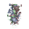 4uw7SC 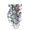 5aq5C S: Starting model for refinement C: citing same article ( |
|---|---|
| Similar structure data |


-Links
 PDBj
PDBj


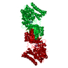
- Assembly
| Deposited unit | 
| |||||||||||||||||||||||||||||||||||||||||||||||||||||||||||||||||||||||||||||||||||||||||||||||||||||||||||||||||||||||||||||||||||||||||||||||||||||||||||||||||||||||||||||||||||||||||||||
|---|---|---|---|---|---|---|---|---|---|---|---|---|---|---|---|---|---|---|---|---|---|---|---|---|---|---|---|---|---|---|---|---|---|---|---|---|---|---|---|---|---|---|---|---|---|---|---|---|---|---|---|---|---|---|---|---|---|---|---|---|---|---|---|---|---|---|---|---|---|---|---|---|---|---|---|---|---|---|---|---|---|---|---|---|---|---|---|---|---|---|---|---|---|---|---|---|---|---|---|---|---|---|---|---|---|---|---|---|---|---|---|---|---|---|---|---|---|---|---|---|---|---|---|---|---|---|---|---|---|---|---|---|---|---|---|---|---|---|---|---|---|---|---|---|---|---|---|---|---|---|---|---|---|---|---|---|---|---|---|---|---|---|---|---|---|---|---|---|---|---|---|---|---|---|---|---|---|---|---|---|---|---|---|---|---|---|---|---|---|---|
| 1 | 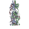
| |||||||||||||||||||||||||||||||||||||||||||||||||||||||||||||||||||||||||||||||||||||||||||||||||||||||||||||||||||||||||||||||||||||||||||||||||||||||||||||||||||||||||||||||||||||||||||||
| 2 | 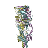
| |||||||||||||||||||||||||||||||||||||||||||||||||||||||||||||||||||||||||||||||||||||||||||||||||||||||||||||||||||||||||||||||||||||||||||||||||||||||||||||||||||||||||||||||||||||||||||||
| 3 | 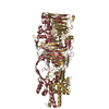
| |||||||||||||||||||||||||||||||||||||||||||||||||||||||||||||||||||||||||||||||||||||||||||||||||||||||||||||||||||||||||||||||||||||||||||||||||||||||||||||||||||||||||||||||||||||||||||||
| Unit cell |
| |||||||||||||||||||||||||||||||||||||||||||||||||||||||||||||||||||||||||||||||||||||||||||||||||||||||||||||||||||||||||||||||||||||||||||||||||||||||||||||||||||||||||||||||||||||||||||||
| Components on special symmetry positions |
| |||||||||||||||||||||||||||||||||||||||||||||||||||||||||||||||||||||||||||||||||||||||||||||||||||||||||||||||||||||||||||||||||||||||||||||||||||||||||||||||||||||||||||||||||||||||||||||
| Noncrystallographic symmetry (NCS) | NCS domain:
|

