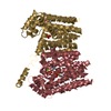
 Movie
Movie Controller


+ Open data

- Basic information
| Entry | Database: PDB / ID: 4u7p | ||||||
|---|---|---|---|---|---|---|---|
| Title | Crystal structure of DNMT3A-DNMT3L complex | ||||||
 Components Components |
| ||||||
 Keywords Keywords | TRANSFERASE/TRANSFERASE REGULATOR / DNA methyltransferase / autoinhibitory form / TRANSFERASE-TRANSFERASE REGULATOR complex | ||||||
| Function / homology |  Function and homology information Function and homology informationepigenetic programing of female pronucleus / chorionic trophoblast cell differentiation / negative regulation of DNA methylation-dependent heterochromatin formation / transposable element silencing by heterochromatin formation / transposable element silencing by piRNA-mediated DNA methylation / protein-cysteine methyltransferase activity / positive regulation of cellular response to hypoxia / cellular response to bisphenol A / regulatory ncRNA-mediated heterochromatin formation / unmethylated CpG binding ...epigenetic programing of female pronucleus / chorionic trophoblast cell differentiation / negative regulation of DNA methylation-dependent heterochromatin formation / transposable element silencing by heterochromatin formation / transposable element silencing by piRNA-mediated DNA methylation / protein-cysteine methyltransferase activity / positive regulation of cellular response to hypoxia / cellular response to bisphenol A / regulatory ncRNA-mediated heterochromatin formation / unmethylated CpG binding / DNA (cytosine-5-)-methyltransferase / genomic imprinting / DNA (cytosine-5-)-methyltransferase activity / autosome genomic imprinting / ESC/E(Z) complex / SUMOylation of DNA methylation proteins / XY body / oocyte development / response to vitamin A / DNA methylation-dependent constitutive heterochromatin formation / response to ionizing radiation / hepatocyte apoptotic process / negative regulation of gene expression via chromosomal CpG island methylation / negative regulation of gene expression, epigenetic / lncRNA binding / male meiosis I / cellular response to ethanol / chromosome, centromeric region / catalytic complex / heterochromatin / placenta development / Transferases; Transferring one-carbon groups; Methyltransferases / DNA methylation / stem cell differentiation / condensed nuclear chromosome / post-embryonic development / PRC2 methylates histones and DNA / Defective pyroptosis / response to cocaine / cellular response to amino acid stimulus / Regulation of endogenous retroelements by Piwi-interacting RNAs (piRNAs) / euchromatin / response to lead ion / response to toxic substance / enzyme activator activity / neuron differentiation / nuclear matrix / RMTs methylate histone arginines / transcription corepressor activity / response to estradiol / methylation / spermatogenesis / cellular response to hypoxia / RNA polymerase II-specific DNA-binding transcription factor binding / RNA polymerase II cis-regulatory region sequence-specific DNA binding / response to xenobiotic stimulus / negative regulation of DNA-templated transcription / chromatin binding / enzyme binding / negative regulation of transcription by RNA polymerase II / DNA binding / zinc ion binding / nucleoplasm / identical protein binding / nucleus / cytoplasm / cytosol Similarity search - Function | ||||||
| Biological species |  Homo sapiens (human) Homo sapiens (human) | ||||||
| Method |  X-RAY DIFFRACTION / SYNCHROTRON / MOLECULAR REPLACEMENT / molecular replacement / Resolution: 3.821 Å X-RAY DIFFRACTION / SYNCHROTRON / MOLECULAR REPLACEMENT / molecular replacement / Resolution: 3.821 Å | ||||||
 Authors Authors | Wang, L. / Guo, X. / Li, J. / Xiao, J. / Yin, X. / He, S. / Wang, J. / Xu, Y. | ||||||
 Citation Citation | Journal: Nature / Year: 2015 Title: Structural insight into autoinhibition and histone H3-induced activation of DNMT3A Authors: Guo, X. / Wang, L. / Li, J. / Ding, Z. / Xiao, J. / Yin, X. / He, S. / Shi, P. / Dong, L. / Li, G. / Tian, C. / Wang, J. / Cong, Y. / Xu, Y. | ||||||
| History |
|
- Structure visualization
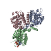
| Structure viewer | Molecule: MolmilJmol/JSmol |
|---|
- Downloads & links
-Download
| PDBx/mmCIF format | 4u7p.cif.gz | 144.8 KB | Display | PDBx/mmCIF format |
|---|---|---|---|---|
| PDB format | pdb4u7p.ent.gz | 109.6 KB | Display | PDB format |
| PDBx/mmJSON format | 4u7p.json.gz | Tree view | PDBx/mmJSON format | |
| Others |  Other downloads Other downloads |
-Validation report
| Arichive directory | https://data.pdbj.org/pub/pdb/validation_reports/u7/4u7pftp://data.pdbj.org/pub/pdb/validation_reports/u7/4u7p | HTTPS FTP |
|---|
-Related structure data
| Related structure data |  4u7tC  2qrvS 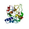 3a1aS C: citing same article ( S: Starting model for refinement |
|---|---|
| Similar structure data |


-Links
 PDBj
PDBj

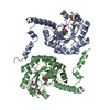
- Assembly
| Deposited unit | 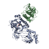
| ||||||||
|---|---|---|---|---|---|---|---|---|---|
| 1 |
| ||||||||
| Unit cell |
| ||||||||
| Details | biological unit is the same as asym. |
-Components
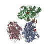
| #1: Protein | Mass: 53350.352 Da / Num. of mol.: 1 / Fragment: UNP residues 455-912 Source method: isolated from a genetically manipulated source Source: (gene. exp.) Homo sapiens (human) / Gene: DNMT3A / Production host:  References: UniProt: Q9Y6K1, DNA (cytosine-5-)-methyltransferase | ||
|---|---|---|---|
| #2: Protein | Mass: 24119.654 Da / Num. of mol.: 1 / Fragment: UNP residues 178-379 Source method: isolated from a genetically manipulated source Source: (gene. exp.) Homo sapiens (human) / Gene: DNMT3L / Production host: | ||
| #3: Chemical |   Mass: 65.409 Da / Num. of mol.: 3 / Source method: obtained synthetically / Formula: Zn Mass: 65.409 Da / Num. of mol.: 3 / Source method: obtained synthetically / Formula: Zn#4: Chemical | ChemComp-SAH / |   Type: L-peptide linking / Mass: 384.411 Da / Num. of mol.: 1 / Source method: obtained synthetically / Formula: C14H20N6O5S Type: L-peptide linking / Mass: 384.411 Da / Num. of mol.: 1 / Source method: obtained synthetically / Formula: C14H20N6O5S |
-Experimental details
-Experiment
| Experiment | Method: X-RAY DIFFRACTION / Number of used crystals: 1 |
|---|
- Sample preparation
| Crystal | Density Matthews: 4.46 Å3/Da / Density % sol: 72.41 % |
|---|---|
| Crystal grow | Temperature: 291 K / Method: vapor diffusion / pH: 5.6 / Details: 0.05M Bis-Tris, 0.1M Sodium Malonate, 8% PEG3350 / PH range: 5.6-6.0 |
-Data collection
| Diffraction | Mean temperature: 100 K | |||||||||||||||||||||||||||||||||||||||||||||||||||||||||||||||||||||||||||||||||||||||||||||||||||
|---|---|---|---|---|---|---|---|---|---|---|---|---|---|---|---|---|---|---|---|---|---|---|---|---|---|---|---|---|---|---|---|---|---|---|---|---|---|---|---|---|---|---|---|---|---|---|---|---|---|---|---|---|---|---|---|---|---|---|---|---|---|---|---|---|---|---|---|---|---|---|---|---|---|---|---|---|---|---|---|---|---|---|---|---|---|---|---|---|---|---|---|---|---|---|---|---|---|---|---|---|
| Diffraction source | Source: SYNCHROTRON / Site: SSRF  / Beamline: BL17U / Wavelength: 1.2816 Å / Beamline: BL17U / Wavelength: 1.2816 Å | |||||||||||||||||||||||||||||||||||||||||||||||||||||||||||||||||||||||||||||||||||||||||||||||||||
| Detector | Type: ADSC QUANTUM 315r / Detector: CCD / Date: Nov 10, 2013 | |||||||||||||||||||||||||||||||||||||||||||||||||||||||||||||||||||||||||||||||||||||||||||||||||||
| Radiation | Monochromator: sagitally focused Si(111) / Protocol: SINGLE WAVELENGTH / Monochromatic (M) / Laue (L): M / Scattering type: x-ray | |||||||||||||||||||||||||||||||||||||||||||||||||||||||||||||||||||||||||||||||||||||||||||||||||||
| Radiation wavelength | Wavelength: 1.2816 Å / Relative weight: 1 | |||||||||||||||||||||||||||||||||||||||||||||||||||||||||||||||||||||||||||||||||||||||||||||||||||
| Reflection | Redundancy: 5.7 % / Number: 80080 / Rmerge(I) obs: 0.098 / Χ2: 0.88 / D res high: 3.82 Å / D res low: 50 Å / Num. obs: 14153 / % possible obs: 99.9 | |||||||||||||||||||||||||||||||||||||||||||||||||||||||||||||||||||||||||||||||||||||||||||||||||||
| Diffraction reflection shell |
| |||||||||||||||||||||||||||||||||||||||||||||||||||||||||||||||||||||||||||||||||||||||||||||||||||
| Reflection | Resolution: 3.82→50 Å / Num. obs: 14153 / % possible obs: 99.9 % / Redundancy: 5.7 % / Biso Wilson estimate: 46.31 Å2 / Rmerge(I) obs: 0.098 / Rpim(I) all: 0.046 / Rrim(I) all: 0.108 / Χ2: 0.885 / Net I/av σ(I): 13.222 / Net I/σ(I): 6.9 / Num. measured all: 80080 | |||||||||||||||||||||||||||||||||||||||||||||||||||||||||||||||||||||||||||||||||||||||||||||||||||
| Reflection shell | Diffraction-ID: 1 / Rejects: _
|
-Phasing
| Phasing | Method: molecular replacement |
|---|
- Processing
| Software |
| ||||||||||||||||||||||||||||||||||||||||||||||||||||||||||||||||||||||||||||||||||||||||||||||||||||||||||||||||
|---|---|---|---|---|---|---|---|---|---|---|---|---|---|---|---|---|---|---|---|---|---|---|---|---|---|---|---|---|---|---|---|---|---|---|---|---|---|---|---|---|---|---|---|---|---|---|---|---|---|---|---|---|---|---|---|---|---|---|---|---|---|---|---|---|---|---|---|---|---|---|---|---|---|---|---|---|---|---|---|---|---|---|---|---|---|---|---|---|---|---|---|---|---|---|---|---|---|---|---|---|---|---|---|---|---|---|---|---|---|---|---|---|---|
| Refinement | Method to determine structure: MOLECULAR REPLACEMENT Starting model: 2QRV, 3A1A Resolution: 3.821→42.005 Å / SU ML: 0.45 / Cross valid method: FREE R-VALUE / σ(F): 1.33 / Phase error: 25.85 / Stereochemistry target values: ML
| ||||||||||||||||||||||||||||||||||||||||||||||||||||||||||||||||||||||||||||||||||||||||||||||||||||||||||||||||
| Solvent computation | Shrinkage radii: 0.9 Å / VDW probe radii: 1.11 Å / Solvent model: FLAT BULK SOLVENT MODEL | ||||||||||||||||||||||||||||||||||||||||||||||||||||||||||||||||||||||||||||||||||||||||||||||||||||||||||||||||
| Displacement parameters | Biso max: 431.61 Å2 / Biso mean: 78.3298 Å2 / Biso min: 0 Å2 | ||||||||||||||||||||||||||||||||||||||||||||||||||||||||||||||||||||||||||||||||||||||||||||||||||||||||||||||||
| Refinement step | Cycle: final / Resolution: 3.821→42.005 Å
| ||||||||||||||||||||||||||||||||||||||||||||||||||||||||||||||||||||||||||||||||||||||||||||||||||||||||||||||||
| Refine LS restraints |
| ||||||||||||||||||||||||||||||||||||||||||||||||||||||||||||||||||||||||||||||||||||||||||||||||||||||||||||||||
| LS refinement shell | Refine-ID: X-RAY DIFFRACTION / Total num. of bins used: 15
|
