 Movie
Movie Controller

[English] 日本語
 Yorodumi
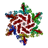
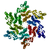
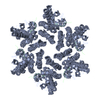
- PDB-4u0a: Hexameric HIV-1 CA in complex with CPSF6 peptide, P6 crystal form -
+ Open data

- Basic information
| Entry | Database: PDB / ID: 4u0a | ||||||
|---|---|---|---|---|---|---|---|
| Title | Hexameric HIV-1 CA in complex with CPSF6 peptide, P6 crystal form | ||||||
 Components Components |
| ||||||
 Keywords Keywords | VIRAL PROTEIN / Capsid | ||||||
| Function / homology |  Function and homology information Function and homology informationexon-exon junction complex binding / positive regulation of RNA export from nucleus / mRNA cleavage factor complex / co-transcriptional mRNA 3'-end processing, cleavage and polyadenylation pathway / interchromatin granule / perichromatin fibrils / Processing of Intronless Pre-mRNAs / mRNA alternative polyadenylation / mRNA cleavage and polyadenylation specificity factor complex / mRNA 3'-end processing ...exon-exon junction complex binding / positive regulation of RNA export from nucleus / mRNA cleavage factor complex / co-transcriptional mRNA 3'-end processing, cleavage and polyadenylation pathway / interchromatin granule / perichromatin fibrils / Processing of Intronless Pre-mRNAs / mRNA alternative polyadenylation / mRNA cleavage and polyadenylation specificity factor complex / mRNA 3'-end processing / Signaling by cytosolic FGFR1 fusion mutants / paraspeckles / mRNA 3'-end processing / RNA Polymerase II Transcription Termination / protein heterotetramerization / viral budding via host ESCRT complex / ribosomal large subunit binding / Processing of Capped Intron-Containing Pre-mRNA / Signaling by FGFR1 in disease / protein tetramerization / host multivesicular body / ISG15 antiviral mechanism / mRNA processing / viral nucleocapsid / nuclear speck / ribonucleoprotein complex / viral translational frameshifting / mRNA binding / host cell nucleus / host cell plasma membrane / virion membrane / structural molecule activity / RNA binding / zinc ion binding / nucleoplasm / membrane / nucleus / cytoplasm Similarity search - Function | ||||||
| Biological species |  Human immunodeficiency virus type 1 group M subtype B Human immunodeficiency virus type 1 group M subtype B Homo sapiens (human) Homo sapiens (human) | ||||||
| Method |  X-RAY DIFFRACTION / SYNCHROTRON / MOLECULAR REPLACEMENT / molecular replacement / Resolution: 2.05 Å X-RAY DIFFRACTION / SYNCHROTRON / MOLECULAR REPLACEMENT / molecular replacement / Resolution: 2.05 Å | ||||||
 Authors Authors | Price, A.J. / Jacques, D.A. / James, L.C. | ||||||
 Citation Citation | Journal: Plos Pathog. / Year: 2014 Title: Host Cofactors and Pharmacologic Ligands Share an Essential Interface in HIV-1 Capsid That Is Lost upon Disassembly. Authors: Price, A.J. / Jacques, D.A. / McEwan, W.A. / Fletcher, A.J. / Essig, S. / Chin, J.W. / Halambage, U.D. / Aiken, C. / James, L.C. | ||||||
| History |
|
- Structure visualization
| Structure viewer | Molecule: MolmilJmol/JSmol |
|---|
- Downloads & links
-Download
| PDBx/mmCIF format | 4u0a.cif.gz | 101.5 KB | Display | PDBx/mmCIF format |
|---|---|---|---|---|
| PDB format | pdb4u0a.ent.gz | 76.7 KB | Display | PDB format |
| PDBx/mmJSON format | 4u0a.json.gz | Tree view | PDBx/mmJSON format | |
| Others |  Other downloads Other downloads |
-Validation report
| Arichive directory | https://data.pdbj.org/pub/pdb/validation_reports/u0/4u0aftp://data.pdbj.org/pub/pdb/validation_reports/u0/4u0a | HTTPS FTP |
|---|
-Related structure data
| Related structure data | 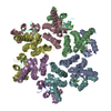 4u0bC  4u0cC  4u0dC 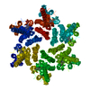 4u0eC  4u0fC  3h47S S: Starting model for refinement C: citing same article ( |
|---|---|
| Similar structure data |


-Links
 PDBj
PDBj


- Assembly
| Deposited unit | 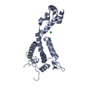
| ||||||||
|---|---|---|---|---|---|---|---|---|---|
| 1 | x 6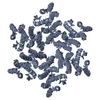
| ||||||||
| Unit cell |
| ||||||||
| Components on special symmetry positions |
| ||||||||
| Details | biological unit is the same as asym. |
-Components
| #1: Protein | Mass: 25461.271 Da / Num. of mol.: 1 / Mutation: A14C,E45C,W184A,M185A Source method: isolated from a genetically manipulated source Source: (gene. exp.) Human immunodeficiency virus type 1 group M subtype BStrain: isolate NY5 / Gene: gag / Plasmid: pET11a / Production host:  |
|---|---|
| #2: Protein/peptide | Mass: 1550.796 Da / Num. of mol.: 1 / Fragment: UNP residues 313-327 / Source method: obtained synthetically / Source: (synth.) Homo sapiens (human) / References: UniProt: Q16630 |
| #3: Chemical | ChemComp-CL /   Mass: 35.453 Da / Num. of mol.: 1 / Source method: obtained synthetically / Formula: Cl Mass: 35.453 Da / Num. of mol.: 1 / Source method: obtained synthetically / Formula: Cl |
| #4: Water | ChemComp-HOH /  Mass: 18.015 Da / Num. of mol.: 147 / Source method: isolated from a natural source / Formula: H2O Mass: 18.015 Da / Num. of mol.: 147 / Source method: isolated from a natural source / Formula: H2O |
| Has protein modification | Y |
-Experimental details
-Experiment
| Experiment | Method: X-RAY DIFFRACTION / Number of used crystals: 1 |
|---|
- Sample preparation
| Crystal | Density Matthews: 2.55 Å3/Da / Density % sol: 51.82 % |
|---|---|
| Crystal grow | Temperature: 290 K / Method: vapor diffusion, sitting drop / pH: 8.5 Details: 0.6M sodium potassium tartrate tetrahydrate, 0.1M TRIS |
-Data collection
| Diffraction | Mean temperature: 100 K | ||||||||||||||||||||||||||||||||||||||||||||||||||||||||||||||||||||||||||||||||||||||||||||||||||||||||||||||
|---|---|---|---|---|---|---|---|---|---|---|---|---|---|---|---|---|---|---|---|---|---|---|---|---|---|---|---|---|---|---|---|---|---|---|---|---|---|---|---|---|---|---|---|---|---|---|---|---|---|---|---|---|---|---|---|---|---|---|---|---|---|---|---|---|---|---|---|---|---|---|---|---|---|---|---|---|---|---|---|---|---|---|---|---|---|---|---|---|---|---|---|---|---|---|---|---|---|---|---|---|---|---|---|---|---|---|---|---|---|---|---|
| Diffraction source | Source: SYNCHROTRON / Site: Diamond  / Beamline: I24 / Wavelength: 0.9686 Å / Beamline: I24 / Wavelength: 0.9686 Å | ||||||||||||||||||||||||||||||||||||||||||||||||||||||||||||||||||||||||||||||||||||||||||||||||||||||||||||||
| Detector | Type: PSI PILATUS 6M / Detector: PIXEL / Date: Jul 6, 2012 | ||||||||||||||||||||||||||||||||||||||||||||||||||||||||||||||||||||||||||||||||||||||||||||||||||||||||||||||
| Radiation | Protocol: SINGLE WAVELENGTH / Monochromatic (M) / Laue (L): M / Scattering type: x-ray | ||||||||||||||||||||||||||||||||||||||||||||||||||||||||||||||||||||||||||||||||||||||||||||||||||||||||||||||
| Radiation wavelength | Wavelength: 0.9686 Å / Relative weight: 1 | ||||||||||||||||||||||||||||||||||||||||||||||||||||||||||||||||||||||||||||||||||||||||||||||||||||||||||||||
| Reflection | Resolution: 2.05→79.284 Å / Num. all: 17251 / Num. obs: 17251 / % possible obs: 100 % / Redundancy: 7.3 % / Rsym value: 0.266 / Net I/σ(I): 4.6 | ||||||||||||||||||||||||||||||||||||||||||||||||||||||||||||||||||||||||||||||||||||||||||||||||||||||||||||||
| Reflection shell | Diffraction-ID: 1 / Rejects: _
|
-Phasing
| Phasing | Method: molecular replacement | |||||||||
|---|---|---|---|---|---|---|---|---|---|---|
| Phasing MR | Model details: Phaser MODE: MR_AUTO
|
- Processing
| Software |
| |||||||||||||||||||||||||||||||||||||||||||||||||||||||||||||||||||||||||||
|---|---|---|---|---|---|---|---|---|---|---|---|---|---|---|---|---|---|---|---|---|---|---|---|---|---|---|---|---|---|---|---|---|---|---|---|---|---|---|---|---|---|---|---|---|---|---|---|---|---|---|---|---|---|---|---|---|---|---|---|---|---|---|---|---|---|---|---|---|---|---|---|---|---|---|---|---|
| Refinement | Method to determine structure: MOLECULAR REPLACEMENT Starting model: PDB entry 3H47 Resolution: 2.05→79.28 Å / Cor.coef. Fo:Fc: 0.924 / Cor.coef. Fo:Fc free: 0.899 / WRfactor Rfree: 0.2656 / WRfactor Rwork: 0.2259 / FOM work R set: 0.8054 / SU B: 10.218 / SU ML: 0.141 / SU R Cruickshank DPI: 0.2041 / SU Rfree: 0.1785 / Cross valid method: THROUGHOUT / σ(F): 0 / ESU R: 0.204 / ESU R Free: 0.179 / Stereochemistry target values: MAXIMUM LIKELIHOOD Details: HYDROGENS HAVE BEEN ADDED IN THE RIDING POSITIONS U VALUES : WITH TLS ADDED
| |||||||||||||||||||||||||||||||||||||||||||||||||||||||||||||||||||||||||||
| Solvent computation | Ion probe radii: 0.8 Å / Shrinkage radii: 0.8 Å / VDW probe radii: 1.2 Å / Solvent model: MASK | |||||||||||||||||||||||||||||||||||||||||||||||||||||||||||||||||||||||||||
| Displacement parameters | Biso max: 79.22 Å2 / Biso mean: 27.74 Å2 / Biso min: 12.44 Å2
| |||||||||||||||||||||||||||||||||||||||||||||||||||||||||||||||||||||||||||
| Refinement step | Cycle: final / Resolution: 2.05→79.28 Å
| |||||||||||||||||||||||||||||||||||||||||||||||||||||||||||||||||||||||||||
| Refine LS restraints |
| |||||||||||||||||||||||||||||||||||||||||||||||||||||||||||||||||||||||||||
| LS refinement shell | Resolution: 2.05→2.103 Å / Total num. of bins used: 20
| |||||||||||||||||||||||||||||||||||||||||||||||||||||||||||||||||||||||||||
| Refinement TLS params. | Method: refined / Refine-ID: X-RAY DIFFRACTION
| |||||||||||||||||||||||||||||||||||||||||||||||||||||||||||||||||||||||||||
| Refinement TLS group |
|
