 Movie
Movie Controller


+ Open data

- Basic information
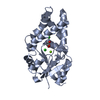
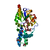
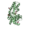
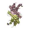
| Entry | Database: PDB / ID: 4twi | ||||||
|---|---|---|---|---|---|---|---|
| Title | The structure of Sir2Af1 bound to a succinylated histone peptide | ||||||
 Components Components |
| ||||||
 Keywords Keywords | HYDROLASE / Sirtuin / desuccinylation / archaeal proteins / histone peptide | ||||||
| Function / homology |  Function and homology information Function and homology informationprotein-malonyllysine demalonylase activity / protein-succinyllysine desuccinylase activity / HATs acetylate histones / RNA polymerase I upstream activating factor complex / Condensation of Prophase Chromosomes / : / : / : / Assembly of the ORC complex at the origin of replication / HDACs deacetylate histones ...protein-malonyllysine demalonylase activity / protein-succinyllysine desuccinylase activity / HATs acetylate histones / RNA polymerase I upstream activating factor complex / Condensation of Prophase Chromosomes / : / : / : / Assembly of the ORC complex at the origin of replication / HDACs deacetylate histones / protein acetyllysine N-acetyltransferase / histone deacetylase activity, NAD-dependent / Recruitment and ATM-mediated phosphorylation of repair and signaling proteins at DNA double strand breaks / Oxidative Stress Induced Senescence / RMTs methylate histone arginines / SUMOylation of chromatin organization proteins / RNA Polymerase I Promoter Escape / positive regulation of transcription by RNA polymerase I / nucleolar large rRNA transcription by RNA polymerase I / Estrogen-dependent gene expression / NAD+ binding / structural constituent of chromatin / nucleosome / nucleosome assembly / chromatin organization / protein heterodimerization activity / regulation of DNA-templated transcription / DNA binding / zinc ion binding / identical protein binding / nucleus / cytoplasm Similarity search - Function | ||||||
| Biological species |   Archaeoglobus fulgidus (archaea) Archaeoglobus fulgidus (archaea) | ||||||
| Method |  X-RAY DIFFRACTION / MOLECULAR REPLACEMENT / Resolution: 1.79 Å X-RAY DIFFRACTION / MOLECULAR REPLACEMENT / Resolution: 1.79 Å | ||||||
 Authors Authors | Ringel, A.E. / Roman, C. / Wolberger, C. | ||||||
| Funding support |  United States, 1items United States, 1items
| ||||||
 Citation Citation | Journal: Protein Sci. / Year: 2014 Title: Alternate deacylating specificities of the archaeal sirtuins Sir2Af1 and Sir2Af2. Authors: Ringel, A.E. / Roman, C. / Wolberger, C. | ||||||
| History |
|
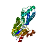
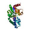
- Structure visualization
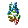
| Structure viewer | Molecule: MolmilJmol/JSmol |
|---|
- Downloads & links
-Download
| PDBx/mmCIF format | 4twi.cif.gz | 71.3 KB | Display | PDBx/mmCIF format |
|---|---|---|---|---|
| PDB format | pdb4twi.ent.gz | 49.3 KB | Display | PDB format |
| PDBx/mmJSON format | 4twi.json.gz | Tree view | PDBx/mmJSON format | |
| Others |  Other downloads Other downloads |
-Validation report
| Arichive directory | https://data.pdbj.org/pub/pdb/validation_reports/tw/4twiftp://data.pdbj.org/pub/pdb/validation_reports/tw/4twi | HTTPS FTP |
|---|
-Related structure data
| Related structure data | 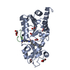 4twjC 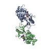 1iciS C: citing same article ( S: Starting model for refinement |
|---|---|
| Similar structure data |


-Links
 PDBj
PDBj


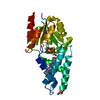
- Assembly
| Deposited unit | 
| ||||||||
|---|---|---|---|---|---|---|---|---|---|
| 1 |
| ||||||||
| Unit cell |
|
-Components
| #1: Protein | Mass: 27181.408 Da / Num. of mol.: 1 Source method: isolated from a genetically manipulated source Source: (gene. exp.) Archaeoglobus fulgidus (archaea)Strain: ATCC 49558 / VC-16 / DSM 4304 / JCM 9628 / NBRC 100126 Gene: cobB1, AF_1676 / Plasmid: pET11a / Production host:  References: UniProt: O28597, Hydrolases; Acting on carbon-nitrogen bonds, other than peptide bonds; In linear amides |
|---|---|
| #2: Protein/peptide | Mass: 1684.986 Da / Num. of mol.: 1 / Fragment: UNP residues 9-21 / Source method: obtained synthetically / Source: (synth.) |
| #3: Chemical | ChemComp-ZN /   Mass: 65.409 Da / Num. of mol.: 1 / Source method: obtained synthetically / Formula: Zn Mass: 65.409 Da / Num. of mol.: 1 / Source method: obtained synthetically / Formula: Zn |
| #4: Chemical | ChemComp-GOL /   Mass: 92.094 Da / Num. of mol.: 1 / Source method: obtained synthetically / Formula: C3H8O3 Mass: 92.094 Da / Num. of mol.: 1 / Source method: obtained synthetically / Formula: C3H8O3 |
| #5: Water | ChemComp-HOH /  Mass: 18.015 Da / Num. of mol.: 238 / Source method: isolated from a natural source / Formula: H2O Mass: 18.015 Da / Num. of mol.: 238 / Source method: isolated from a natural source / Formula: H2O |
-Experimental details
-Experiment
| Experiment | Method: X-RAY DIFFRACTION |
|---|
- Sample preparation
| Crystal | Density Matthews: 2.24 Å3/Da / Density % sol: 45.11 % |
|---|---|
| Crystal grow | Temperature: 293 K / Method: vapor diffusion, hanging drop / pH: 8.5 Details: 0.1 M Tris pH 8.5, 200 mM MgCl2, and 20% (w/v) PEG 8,000 |
-Data collection
| Diffraction | Mean temperature: 100 K |
|---|---|
| Diffraction source | Source: ROTATING ANODE / Type: RIGAKU FR-E SUPERBRIGHT / Wavelength: 1.54 Å |
| Detector | Type: RIGAKU SATURN 944+ / Detector: CCD / Date: Jan 10, 2014 |
| Radiation | Protocol: SINGLE WAVELENGTH / Monochromatic (M) / Laue (L): M / Scattering type: x-ray |
| Radiation wavelength | Wavelength: 1.54 Å / Relative weight: 1 |
| Reflection | Resolution: 1.79→21.94 Å / Num. all: 53220 / Num. obs: 23269 / % possible obs: 95.39 % / Redundancy: 2.3 % / Rsym value: 0.1172 / Net I/σ(I): 7.36 |
| Reflection shell | Resolution: 1.79→1.849 Å / Redundancy: 1.5 % / Mean I/σ(I) obs: 1.85 / % possible all: 83.73 |
- Processing
| Software | Name: REFMAC / Version: 5.7.0029 / Classification: refinement | ||||||||||||||||||||||||||||||||||||||||||||||||||||||||||||||||||||||||||||||||||||||||||||||||||||||||||||||||||||||||||||||||||||||||||||||||||||||||||||||||||||||||||||||||||||||
|---|---|---|---|---|---|---|---|---|---|---|---|---|---|---|---|---|---|---|---|---|---|---|---|---|---|---|---|---|---|---|---|---|---|---|---|---|---|---|---|---|---|---|---|---|---|---|---|---|---|---|---|---|---|---|---|---|---|---|---|---|---|---|---|---|---|---|---|---|---|---|---|---|---|---|---|---|---|---|---|---|---|---|---|---|---|---|---|---|---|---|---|---|---|---|---|---|---|---|---|---|---|---|---|---|---|---|---|---|---|---|---|---|---|---|---|---|---|---|---|---|---|---|---|---|---|---|---|---|---|---|---|---|---|---|---|---|---|---|---|---|---|---|---|---|---|---|---|---|---|---|---|---|---|---|---|---|---|---|---|---|---|---|---|---|---|---|---|---|---|---|---|---|---|---|---|---|---|---|---|---|---|---|---|
| Refinement | Method to determine structure: MOLECULAR REPLACEMENT Starting model: 1ICI Resolution: 1.79→21.94 Å / Cor.coef. Fo:Fc: 0.947 / Cor.coef. Fo:Fc free: 0.932 / SU B: 2.662 / SU ML: 0.084 / Cross valid method: THROUGHOUT / ESU R: 0.145 / ESU R Free: 0.129 / Stereochemistry target values: MAXIMUM LIKELIHOOD / Details: HYDROGENS HAVE BEEN ADDED IN THE RIDING POSITIONS
| ||||||||||||||||||||||||||||||||||||||||||||||||||||||||||||||||||||||||||||||||||||||||||||||||||||||||||||||||||||||||||||||||||||||||||||||||||||||||||||||||||||||||||||||||||||||
| Solvent computation | Ion probe radii: 0.8 Å / Shrinkage radii: 0.8 Å / VDW probe radii: 1.2 Å / Solvent model: MASK | ||||||||||||||||||||||||||||||||||||||||||||||||||||||||||||||||||||||||||||||||||||||||||||||||||||||||||||||||||||||||||||||||||||||||||||||||||||||||||||||||||||||||||||||||||||||
| Displacement parameters | Biso mean: 19.4 Å2
| ||||||||||||||||||||||||||||||||||||||||||||||||||||||||||||||||||||||||||||||||||||||||||||||||||||||||||||||||||||||||||||||||||||||||||||||||||||||||||||||||||||||||||||||||||||||
| Refinement step | Cycle: 1 / Resolution: 1.79→21.94 Å
| ||||||||||||||||||||||||||||||||||||||||||||||||||||||||||||||||||||||||||||||||||||||||||||||||||||||||||||||||||||||||||||||||||||||||||||||||||||||||||||||||||||||||||||||||||||||
| Refine LS restraints |
|