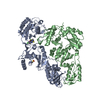
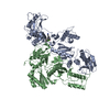
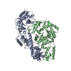
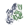
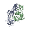
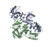
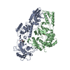
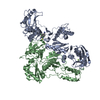
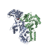
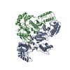
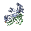
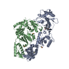
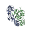
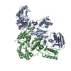
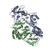
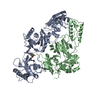
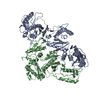
登録情報 データベース : PDB / ID : 4rw8タイトル Crystal Structure of HIV-1 Reverse Transcriptase in complex with (E)-3-(3-chloro-5-(2-(2-(2,4-dioxo-3,4-dihydropyrimidin-1(2H)-yl)ethoxy)phenoxy)phenyl)acrylonitrile (JLJ532), a non-nucleoside inhibitor' Reverse transcriptase/ribonuclease H, p51 subunit Reverse transcriptase/ribonuclease H, p66 subunit キーワード / / / / 機能・相同性 分子機能 ドメイン・相同性 構成要素
/ / / / / / / / / / / / / / / / / / / / / / / / / / / / / / / / / / / / / / / / / / / / / / / / / / / / / / / / / / / / / / / / / / / / / / / / / / / / / / / / / / / / / / / / / / / / / / / / / / / / / / / / 生物種 手法 / / / 解像度 : 2.878 Å データ登録者 Frey, K.M. / Anderson, K.S. ジャーナル : J.Med.Chem. / 年 : 2015タイトル : Structure-Based Evaluation of Non-nucleoside Inhibitors with Improved Potency and Solubility That Target HIV Reverse Transcriptase Variants.著者 : Frey, K.M. / Puleo, D.E. / Spasov, K.A. / Bollini, M. / Jorgensen, W.L. / Anderson, K.S. 履歴 登録 2014年12月1日 登録サイト / 処理サイト 改定 1.0 2015年4月29日 Provider / タイプ 改定 1.1 2017年11月22日 Group / カテゴリ 改定 1.2 2023年9月20日 Group Data collection / Database references ... Data collection / Database references / Derived calculations / Refinement description カテゴリ chem_comp_atom / chem_comp_bond ... chem_comp_atom / chem_comp_bond / database_2 / pdbx_initial_refinement_model / struct_ref_seq_dif / struct_site Item _database_2.pdbx_DOI / _database_2.pdbx_database_accession ... _database_2.pdbx_DOI / _database_2.pdbx_database_accession / _struct_ref_seq_dif.details / _struct_site.pdbx_auth_asym_id / _struct_site.pdbx_auth_comp_id / _struct_site.pdbx_auth_seq_id
すべて表示 表示を減らす
 ムービー
ムービー コントローラー


 データを開く
 基本情報
基本情報 要素
要素 キーワード
キーワード 機能・相同性情報
 Human immunodeficiency virus type 1 BH10 (ヒト免疫不全ウイルス)
 X線回折 /
  データ登録者
 引用
引用 構造の表示
構造の表示 ダウンロードとリンク
 その他のダウンロード


 PDBj
PDBj


 集合体
集合体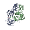

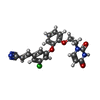
 分子量: 409.822 Da / 分子数: 1 / 由来タイプ: 合成 / 式: C21H16ClN3O4
 分子量: 18.015 Da / 分子数: 19 / 由来タイプ: 天然 / 式: H2O
 試料調製
試料調製 / ビームライン: X29A / 波長: 1.075 Å
 解析
解析