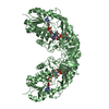
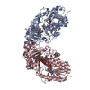
 Movie
Movie Controller

[English] 日本語
 Yorodumi
- PDB-4rpg: Crystal structure of Micobacterium tuberculosis UDP-Galactopyrano... -
+ Open data

- Basic information
| Entry | Database: PDB / ID: 4rpg | ||||||
|---|---|---|---|---|---|---|---|
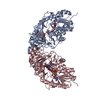
| Title | Crystal structure of Micobacterium tuberculosis UDP-Galactopyranose mutase in complex with substrate UDP-Galp | ||||||
 Components Components | UDP-galactopyranose mutase | ||||||
 Keywords Keywords | ISOMERASE / UDP-galactopyranose mutase / MtUGM / flavoenzyme / FAD | ||||||
| Function / homology |  Function and homology information Function and homology informationUDP-galactopyranose mutase / UDP-galactopyranose mutase activity / Actinobacterium-type cell wall biogenesis / capsule polysaccharide biosynthetic process / peptidoglycan-based cell wall / cell wall organization / flavin adenine dinucleotide binding / plasma membrane / cytosol Similarity search - Function | ||||||
| Biological species |   Mycobacterium tuberculosis (bacteria) Mycobacterium tuberculosis (bacteria) | ||||||
| Method |  X-RAY DIFFRACTION / SYNCHROTRON / MOLECULAR REPLACEMENT / molecular replacement / Resolution: 2.4001 Å X-RAY DIFFRACTION / SYNCHROTRON / MOLECULAR REPLACEMENT / molecular replacement / Resolution: 2.4001 Å | ||||||
 Authors Authors | Van Straaten, K.E. / Sanders, D.A.R. | ||||||
 Citation Citation | Journal: J.Am.Chem.Soc. / Year: 2015 Title: Structural Basis of Ligand Binding to UDP-Galactopyranose Mutase from Mycobacterium tuberculosis Using Substrate and Tetrafluorinated Substrate Analogues. Authors: van Straaten, K.E. / Kuttiyatveetil, J.R. / Sevrain, C.M. / Villaume, S.A. / Jimenez-Barbero, J. / Linclau, B. / Vincent, S.P. / Sanders, D.A. | ||||||
| History |
|
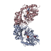
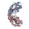
- Structure visualization
| Structure viewer | Molecule: MolmilJmol/JSmol |
|---|
- Downloads & links
-Download
| PDBx/mmCIF format | 4rpg.cif.gz | 261.3 KB | Display | PDBx/mmCIF format |
|---|---|---|---|---|
| PDB format | pdb4rpg.ent.gz | 210.6 KB | Display | PDB format |
| PDBx/mmJSON format | 4rpg.json.gz | Tree view | PDBx/mmJSON format | |
| Others |  Other downloads Other downloads |
-Validation report
| Arichive directory | https://data.pdbj.org/pub/pdb/validation_reports/rp/4rpgftp://data.pdbj.org/pub/pdb/validation_reports/rp/4rpg | HTTPS FTP |
|---|
-Related structure data
| Related structure data |  4rphC  4rpjC  4rpkC  4rplC  1vojS C: citing same article ( S: Starting model for refinement |
|---|---|
| Similar structure data |

-Links
 PDBj
PDBj


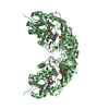
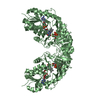
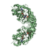
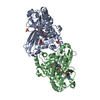
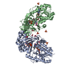
- Assembly
| Deposited unit | 
| ||||||||||||||||||||||||||||||||||||||||
|---|---|---|---|---|---|---|---|---|---|---|---|---|---|---|---|---|---|---|---|---|---|---|---|---|---|---|---|---|---|---|---|---|---|---|---|---|---|---|---|---|---|
| 1 | 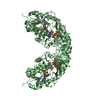
| ||||||||||||||||||||||||||||||||||||||||
| 2 | 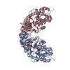
| ||||||||||||||||||||||||||||||||||||||||
| Unit cell |
| ||||||||||||||||||||||||||||||||||||||||
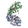
| Noncrystallographic symmetry (NCS) | NCS domain:
NCS domain segments: Component-ID: 1 / Ens-ID: 1 / Beg auth comp-ID: THR / Beg label comp-ID: THR / End auth comp-ID: LEU / End label comp-ID: LEU / Auth seq-ID: 5 - 395 / Label seq-ID: 5 - 395
NCS oper:
|
-Components
| #1: Protein | Mass: 45928.348 Da / Num. of mol.: 3 / Fragment: UDP-Galactopyranose Mutase / Mutation: P306R Source method: isolated from a genetically manipulated source Source: (gene. exp.) Mycobacterium tuberculosis (bacteria) / Strain: ATCC 25618 / H37Rv / Gene: glf, glfA, Rv3809c / Plasmid: pDEST-His-MBP / Production host: #2: Chemical |   Mass: 785.550 Da / Num. of mol.: 3 / Source method: obtained synthetically / Formula: C27H33N9O15P2 / Comment: FAD*YM Mass: 785.550 Da / Num. of mol.: 3 / Source method: obtained synthetically / Formula: C27H33N9O15P2 / Comment: FAD*YM#3: Chemical | ChemComp-GDU / | 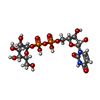  Mass: 566.302 Da / Num. of mol.: 1 Mass: 566.302 Da / Num. of mol.: 1Source method: isolated from a genetically manipulated source Formula: C15H24N2O17P2 #4: Chemical |   Type: RNA linking / Mass: 404.161 Da / Num. of mol.: 2 / Source method: obtained synthetically / Formula: C9H14N2O12P2 / Comment: UDP*YM Type: RNA linking / Mass: 404.161 Da / Num. of mol.: 2 / Source method: obtained synthetically / Formula: C9H14N2O12P2 / Comment: UDP*YM#5: Water | ChemComp-HOH / |  Mass: 18.015 Da / Num. of mol.: 395 / Source method: isolated from a natural source / Formula: H2O Mass: 18.015 Da / Num. of mol.: 395 / Source method: isolated from a natural source / Formula: H2O |
|---|
-Experimental details
-Experiment
| Experiment | Method: X-RAY DIFFRACTION / Number of used crystals: 1 |
|---|
- Sample preparation
| Crystal | Density Matthews: 2.91 Å3/Da / Density % sol: 57.78 % |
|---|---|
| Crystal grow | Temperature: 277 K / Method: hanging drop vapor diffusion / pH: 5.5 Details: 0.1M Bis-Tris pH 5.5, 20% PEG 3350, 10mM Phenol, Hanging drop vapor diffusion, temperature 277K |
-Data collection
| Diffraction | Mean temperature: 100 K |
|---|---|
| Diffraction source | Source: SYNCHROTRON / Site: CLSI  / Beamline: 08ID-1 / Wavelength: 0.97949 Å / Beamline: 08ID-1 / Wavelength: 0.97949 Å |
| Detector | Type: MARMOSAIC 300 mm CCD / Detector: CCD / Date: Jan 10, 2014 |
| Radiation | Protocol: SINGLE WAVELENGTH / Monochromatic (M) / Laue (L): M / Scattering type: x-ray |
| Radiation wavelength | Wavelength: 0.97949 Å / Relative weight: 1 |
| Reflection | Resolution: 2.4→47.3 Å / Num. all: 61216 / Num. obs: 61216 / % possible obs: 98.9 % / Observed criterion σ(F): 0 / Observed criterion σ(I): 0 / Redundancy: 4.2 % / Biso Wilson estimate: 48.2 Å2 / Rmerge(I) obs: 0.094 / Rsym value: 0.094 / Net I/σ(I): 9.68 |
| Reflection shell | Resolution: 2.4→2.46 Å / % possible obs: 98.8 % / Redundancy: 4.1 % / Rmerge(I) obs: 0.764 / Mean I/σ(I) obs: 1.6 / Num. measured all: 4478 / Num. measured obs: 4478 / % possible all: 98.8 |
-Phasing
| Phasing | Method: molecular replacement |
|---|
- Processing
| Software |
| |||||||||||||||||||||||||||||||||||||||||||||||||||||||||||||||||||||||||||||||||||||||||||||||||||||||||||||||||||||||||||||||||||||||||||||||||||
|---|---|---|---|---|---|---|---|---|---|---|---|---|---|---|---|---|---|---|---|---|---|---|---|---|---|---|---|---|---|---|---|---|---|---|---|---|---|---|---|---|---|---|---|---|---|---|---|---|---|---|---|---|---|---|---|---|---|---|---|---|---|---|---|---|---|---|---|---|---|---|---|---|---|---|---|---|---|---|---|---|---|---|---|---|---|---|---|---|---|---|---|---|---|---|---|---|---|---|---|---|---|---|---|---|---|---|---|---|---|---|---|---|---|---|---|---|---|---|---|---|---|---|---|---|---|---|---|---|---|---|---|---|---|---|---|---|---|---|---|---|---|---|---|---|---|---|---|---|
| Refinement | Method to determine structure: MOLECULAR REPLACEMENT Starting model: 1VOJ Resolution: 2.4001→47.27 Å / FOM work R set: 0.7805 / SU ML: 0.37 / Isotropic thermal model: Isotropic / Cross valid method: THROUGHOUT / σ(F): 1.35 / Phase error: 27.93 / Stereochemistry target values: ML
| |||||||||||||||||||||||||||||||||||||||||||||||||||||||||||||||||||||||||||||||||||||||||||||||||||||||||||||||||||||||||||||||||||||||||||||||||||
| Solvent computation | Shrinkage radii: 0.9 Å / VDW probe radii: 1.11 Å / Solvent model: FLAT BULK SOLVENT MODEL | |||||||||||||||||||||||||||||||||||||||||||||||||||||||||||||||||||||||||||||||||||||||||||||||||||||||||||||||||||||||||||||||||||||||||||||||||||
| Displacement parameters | Biso max: 125.63 Å2 / Biso mean: 51.61 Å2 / Biso min: 27.54 Å2 | |||||||||||||||||||||||||||||||||||||||||||||||||||||||||||||||||||||||||||||||||||||||||||||||||||||||||||||||||||||||||||||||||||||||||||||||||||
| Refinement step | Cycle: LAST / Resolution: 2.4001→47.27 Å
| |||||||||||||||||||||||||||||||||||||||||||||||||||||||||||||||||||||||||||||||||||||||||||||||||||||||||||||||||||||||||||||||||||||||||||||||||||
| Refine LS restraints |
| |||||||||||||||||||||||||||||||||||||||||||||||||||||||||||||||||||||||||||||||||||||||||||||||||||||||||||||||||||||||||||||||||||||||||||||||||||
| Refine LS restraints NCS |
| |||||||||||||||||||||||||||||||||||||||||||||||||||||||||||||||||||||||||||||||||||||||||||||||||||||||||||||||||||||||||||||||||||||||||||||||||||
| LS refinement shell | Refine-ID: X-RAY DIFFRACTION / Total num. of bins used: 20
|
