Resolution: 2.5→50 Å / Cor.coef. Fo:Fc: 0.954 / Cor.coef. Fo:Fc free: 0.917 / SU B: 8.933 / SU ML: 0.197 / Cross valid method: THROUGHOUT / σ(F): 0 / ESU R: 0.383 / ESU R Free: 0.26 / Stereochemistry target values: MAXIMUM LIKELIHOOD Details: THE ENTRY CONTAINS FRIEDEL PAIRS IN F_PLUS/MINUS COLUMNS. HYDROGENS HAVE BEEN ADDED IN THE RIDING POSITIONS.
Rfactor
Num. reflection
% reflection
Selection details
Rfree
0.23539
2402
5.1 %
RANDOM
Rwork
0.17188
-
-
-
all
0.17506
-
-
-
obs
0.17506
45052
99.96 %
-
Solvent computation
Ion probe radii: 0.8 Å / Shrinkage radii: 0.8 Å / VDW probe radii: 1.2 Å / Solvent model: MASK
Displacement parameters
Biso mean: 47.757 Å2
Baniso -1
Baniso -2
Baniso -3
1-
0.53 Å2
0 Å2
-0 Å2
2-
-
0.53 Å2
0 Å2
3-
-
-
-1.06 Å2
Refinement step
Cycle: LAST / Resolution: 2.5→50 Å
Protein
Nucleic acid
Ligand
Solvent
Total
Num. atoms
8148
0
149
197
8494
Refine LS restraints
Refine-ID
Type
Dev ideal
Dev ideal target
Number
X-RAY DIFFRACTION
r_bond_refined_d
0.013
0.019
8422
X-RAY DIFFRACTION
r_bond_other_d
0.005
0.02
7627
X-RAY DIFFRACTION
r_angle_refined_deg
1.523
1.96
11386
X-RAY DIFFRACTION
r_angle_other_deg
0.836
3
17619
X-RAY DIFFRACTION
r_dihedral_angle_1_deg
5.638
5
978
X-RAY DIFFRACTION
r_dihedral_angle_2_deg
41.219
25.526
456
X-RAY DIFFRACTION
r_dihedral_angle_3_deg
18.503
15
1506
X-RAY DIFFRACTION
r_dihedral_angle_4_deg
21.564
15
24
X-RAY DIFFRACTION
r_chiral_restr
0.081
0.2
1221
X-RAY DIFFRACTION
r_gen_planes_refined
0.006
0.02
9480
X-RAY DIFFRACTION
r_gen_planes_other
0.001
0.02
1992
X-RAY DIFFRACTION
r_mcbond_it
3.68
4.639
3930
X-RAY DIFFRACTION
r_mcbond_other
3.675
4.638
3929
X-RAY DIFFRACTION
r_mcangle_it
5.76
6.936
4902
X-RAY DIFFRACTION
r_mcangle_other
5.761
6.938
4903
X-RAY DIFFRACTION
r_scbond_it
4.248
4.894
4490
X-RAY DIFFRACTION
r_scbond_other
4.248
4.895
4490
X-RAY DIFFRACTION
r_scangle_other
6.581
7.163
6484
X-RAY DIFFRACTION
r_long_range_B_refined
9.792
36.195
10211
X-RAY DIFFRACTION
r_long_range_B_other
9.789
36.183
10166
LS refinement shell
Resolution: 2.5→2.564 Å / Total num. of bins used: 20
Rfactor
Num. reflection
% reflection
Rfree
0.324
175
-
Rwork
0.249
3314
-
obs
-
-
100 %
+
About Yorodumi
-
News
-
Feb 9, 2022. New format data for meta-information of EMDB entries
New format data for meta-information of EMDB entries
Version 3 of the EMDB header file is now the official format.
The previous official version 1.9 will be removed from the archive.
In the structure databanks used in Yorodumi, some data are registered as the other names, "COVID-19 virus" and "2019-nCoV". Here are the details of the virus and the list of structure data.
Jan 31, 2019. EMDB accession codes are about to change! (news from PDBe EMDB page)
EMDB accession codes are about to change! (news from PDBe EMDB page)
The allocation of 4 digits for EMDB accession codes will soon come to an end. Whilst these codes will remain in use, new EMDB accession codes will include an additional digit and will expand incrementally as the available range of codes is exhausted. The current 4-digit format prefixed with “EMD-” (i.e. EMD-XXXX) will advance to a 5-digit format (i.e. EMD-XXXXX), and so on. It is currently estimated that the 4-digit codes will be depleted around Spring 2019, at which point the 5-digit format will come into force.
The EM Navigator/Yorodumi systems omit the EMD- prefix.
Related info.:Q: What is EMD? / ID/Accession-code notation in Yorodumi/EM Navigator
Yorodumi is a browser for structure data from EMDB, PDB, SASBDB, etc.
This page is also the successor to EM Navigator detail page, and also detail information page/front-end page for Omokage search.
The word "yorodu" (or yorozu) is an old Japanese word meaning "ten thousand". "mi" (miru) is to see.
Related info.:EMDB / PDB / SASBDB / Comparison of 3 databanks / Yorodumi Search / Aug 31, 2016. New EM Navigator & Yorodumi / Yorodumi Papers / Jmol/JSmol / Function and homology information / Changes in new EM Navigator and Yorodumi
 Movie
Movie Controller

 Yorodumi
 Open data
 Basic information
 Components
 Keywords
 Function and homology information
 X-RAY DIFFRACTION /
  Authors
 Citation
 Structure visualization
 Downloads & links
 Other downloads

 PDBj
PDBj


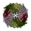
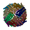
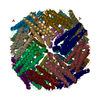
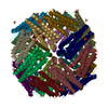
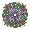
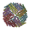
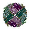
 Assembly


 Mass: 55.845 Da / Num. of mol.: 12 / Source method: obtained synthetically / Formula: Fe
 Mass: 96.063 Da / Num. of mol.: 19 / Source method: obtained synthetically / Formula: SO4
 Mass: 195.237 Da / Num. of mol.: 1 / Source method: obtained synthetically / Formula: C6H13NO4S / Comment: pH buffer*YM
 Mass: 35.453 Da / Num. of mol.: 27 / Source method: obtained synthetically / Formula: Cl
 Mass: 200.590 Da / Num. of mol.: 2 / Source method: obtained synthetically / Formula: Hg
 Mass: 24.305 Da / Num. of mol.: 1 / Source method: obtained synthetically / Formula: Mg
 Sample preparation
 / Beamline: BL26B2 / Wavelength: 1 Å
 Processing