 Movie
Movie Controller

[English] 日本語
 Yorodumi
- PDB-4re3: Different transition state conformations for the hydrolysis of be... -
+ Open data

- Basic information
| Entry | Database: PDB / ID: 4re3 | ||||||
|---|---|---|---|---|---|---|---|
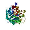
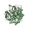
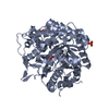
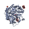
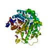
| Title | Different transition state conformations for the hydrolysis of beta-mannosides and beta-glucosides in the rice Os7BGlu26 family GH1 beta-mannosidase/beta-glucosidase | ||||||
 Components Components | Beta-mannosidase/beta-glucosidase | ||||||
 Keywords Keywords | HYDROLASE / GH1 | ||||||
| Function / homology |  Function and homology information Function and homology informationbeta-L-arabinosidase activity / beta-D-fucosidase activity / cellobiose glucosidase activity / beta-mannosidase activity / beta-glucosidase / beta-galactosidase activity / carbohydrate metabolic process Similarity search - Function | ||||||
| Biological species |  | ||||||
| Method |  X-RAY DIFFRACTION / SYNCHROTRON / MOLECULAR REPLACEMENT / Resolution: 2.55 Å X-RAY DIFFRACTION / SYNCHROTRON / MOLECULAR REPLACEMENT / Resolution: 2.55 Å | ||||||
 Authors Authors | Tankrathok, A. / Iglesias-Fernandez, J. / Williams, R.J. / Hakki, Z. / Robinson, R.C. / Hrmova, M. / Rovira, C. / Williams, S.J. / Ketudat Cairns, J.R. | ||||||
 Citation Citation | Journal: ACS CATALYSIS / Year: 2015 Title: A Single Glycosidase Harnesses Different Pyranoside Ring Transition State Conformations for Hydrolysis of Mannosides and Glucosides Authors: Tankrathok, A. / Iglesias-Fernandez, J. / Williams, R.J. / Pengthaisong, S. / Baiya, S. / Hakki, Z. / Robinson, R.C. / Hrmova, M. / Rovira, C. / Williams, S.J. / Ketudat Cairns, J.R. | ||||||
| History |
|
- Structure visualization
| Structure viewer | Molecule: MolmilJmol/JSmol |
|---|
- Downloads & links
-Download
| PDBx/mmCIF format | 4re3.cif.gz | 116.2 KB | Display | PDBx/mmCIF format |
|---|---|---|---|---|
| PDB format | pdb4re3.ent.gz | 87.7 KB | Display | PDB format |
| PDBx/mmJSON format | 4re3.json.gz | Tree view | PDBx/mmJSON format | |
| Others |  Other downloads Other downloads |
-Validation report
| Arichive directory | https://data.pdbj.org/pub/pdb/validation_reports/re/4re3ftp://data.pdbj.org/pub/pdb/validation_reports/re/4re3 | HTTPS FTP |
|---|
-Related structure data
| Related structure data | 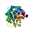 4re2C 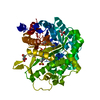 4re4C  4jhoS S: Starting model for refinement C: citing same article ( |
|---|---|
| Similar structure data |


-Links
 PDBj
PDBj- Assembly
| Deposited unit | 
| ||||||||
|---|---|---|---|---|---|---|---|---|---|
| 1 |
| ||||||||
| Unit cell |
|
-Components
-Protein , 1 types, 1 molecules A
| #1: Protein | Mass: 57703.746 Da / Num. of mol.: 1 Source method: isolated from a genetically manipulated source Source: (gene. exp.) Plasmid: pET32A / Production host:  References: UniProt: B5ABY0, UniProt: A2YPH1*PLUS, beta-mannosidase |
|---|
-Non-polymers , 5 types, 126 molecules 


| #2: Chemical | ChemComp-GOL /  Mass: 92.094 Da / Num. of mol.: 4 / Source method: obtained synthetically / Formula: C3H8O3 Mass: 92.094 Da / Num. of mol.: 4 / Source method: obtained synthetically / Formula: C3H8O3#3: Chemical | ChemComp-TRS / |  Mass: 122.143 Da / Num. of mol.: 1 / Source method: obtained synthetically / Formula: C4H12NO3 / Comment: pH buffer*YM Mass: 122.143 Da / Num. of mol.: 1 / Source method: obtained synthetically / Formula: C4H12NO3 / Comment: pH buffer*YM#4: Chemical | ChemComp-EPE / |  Mass: 238.305 Da / Num. of mol.: 1 / Source method: obtained synthetically / Formula: C8H18N2O4S / Comment: pH buffer*YM Mass: 238.305 Da / Num. of mol.: 1 / Source method: obtained synthetically / Formula: C8H18N2O4S / Comment: pH buffer*YM#5: Chemical | ChemComp-GIM / |  Mass: 201.200 Da / Num. of mol.: 1 / Source method: obtained synthetically / Formula: C8H13N2O4 Mass: 201.200 Da / Num. of mol.: 1 / Source method: obtained synthetically / Formula: C8H13N2O4#6: Water | ChemComp-HOH / | Mass: 18.015 Da / Num. of mol.: 119 / Source method: isolated from a natural source / Formula: H2O |
|---|
-Details
| Has protein modification | Y |
|---|
-Experimental details
-Experiment
| Experiment | Method: X-RAY DIFFRACTION / Number of used crystals: 1 |
|---|
- Sample preparation
| Crystal | Density Matthews: 2.97 Å3/Da / Density % sol: 58.63 % |
|---|---|
| Crystal grow | Temperature: 288 K / Method: vapor diffusion, hanging drop / pH: 7.25 Details: 0.8M K/Na tartrate, 0.1 M HEPES, pH 7.25, VAPOR DIFFUSION, HANGING DROP, temperature 288K |
-Data collection
| Diffraction | Mean temperature: 110 K |
|---|---|
| Diffraction source | Source: SYNCHROTRON / Site: NSRRC  / Beamline: BL13B1 / Wavelength: 1 Å / Beamline: BL13B1 / Wavelength: 1 Å |
| Detector | Type: ADSC QUANTUM 315 / Detector: CCD / Date: Dec 1, 2009 |
| Radiation | Protocol: SINGLE WAVELENGTH / Monochromatic (M) / Laue (L): M / Scattering type: x-ray |
| Radiation wavelength | Wavelength: 1 Å / Relative weight: 1 |
| Reflection | Resolution: 2.55→30 Å / Num. all: 22658 / Num. obs: 22658 / % possible obs: 99.9 % / Observed criterion σ(I): 1 |
| Reflection shell | Resolution: 2.55→2.64 Å / % possible all: 100 |
- Processing
| Software |
| ||||||||||||||||||||||||||||||||||||||||||||||||||||||||||||||||||||||||||||||||||||||||||
|---|---|---|---|---|---|---|---|---|---|---|---|---|---|---|---|---|---|---|---|---|---|---|---|---|---|---|---|---|---|---|---|---|---|---|---|---|---|---|---|---|---|---|---|---|---|---|---|---|---|---|---|---|---|---|---|---|---|---|---|---|---|---|---|---|---|---|---|---|---|---|---|---|---|---|---|---|---|---|---|---|---|---|---|---|---|---|---|---|---|---|---|
| Refinement | Method to determine structure: MOLECULAR REPLACEMENT Starting model: 4JHO Resolution: 2.55→24.95 Å / Cor.coef. Fo:Fc: 0.941 / Cor.coef. Fo:Fc free: 0.911 / SU B: 7.318 / SU ML: 0.164 / Cross valid method: THROUGHOUT / ESU R: 0.424 / ESU R Free: 0.243 / Stereochemistry target values: MAXIMUM LIKELIHOOD / Details: HYDROGENS HAVE BEEN ADDED IN THE RIDING POSITIONS
| ||||||||||||||||||||||||||||||||||||||||||||||||||||||||||||||||||||||||||||||||||||||||||
| Solvent computation | Ion probe radii: 0.8 Å / Shrinkage radii: 0 Å / VDW probe radii: 1.2 Å / Solvent model: MASK | ||||||||||||||||||||||||||||||||||||||||||||||||||||||||||||||||||||||||||||||||||||||||||
| Displacement parameters | Biso mean: 20.804 Å2
| ||||||||||||||||||||||||||||||||||||||||||||||||||||||||||||||||||||||||||||||||||||||||||
| Refinement step | Cycle: LAST / Resolution: 2.55→24.95 Å
| ||||||||||||||||||||||||||||||||||||||||||||||||||||||||||||||||||||||||||||||||||||||||||
| Refine LS restraints |
| ||||||||||||||||||||||||||||||||||||||||||||||||||||||||||||||||||||||||||||||||||||||||||
| LS refinement shell | Resolution: 2.545→2.611 Å / Total num. of bins used: 20
|
