 Movie
Movie Controller

[English] 日本語
 Yorodumi
- PDB-4r6d: Modified mTFP* for enhanced metal binding: co-crystallization wit... -
+ Open data

- Basic information
| Entry | Database: PDB / ID: 4r6d | ||||||
|---|---|---|---|---|---|---|---|
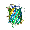
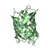
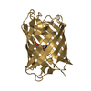
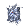
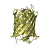
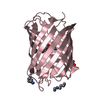
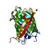
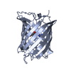
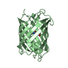
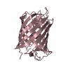
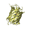
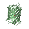
| Title | Modified mTFP* for enhanced metal binding: co-crystallization with CuCl2 | ||||||
 Components Components | GFP-like fluorescent chromoprotein cFP484 | ||||||
 Keywords Keywords | FLUORESCENT PROTEIN / beta barrel topology / engineered metalloenzyme / Diels-Alderase | ||||||
| Function / homology |  Function and homology information Function and homology information | ||||||
| Biological species |  Clavularia sp. (invertebrata) Clavularia sp. (invertebrata) | ||||||
| Method |  X-RAY DIFFRACTION / MOLECULAR REPLACEMENT / Resolution: 1.55 Å X-RAY DIFFRACTION / MOLECULAR REPLACEMENT / Resolution: 1.55 Å | ||||||
 Authors Authors | Fischer, J. / Quitterer, F. / Groll, M. / Eppinger, J. | ||||||
 Citation Citation | Journal: TO BE PUBLISHED Title: Modified mTFP* for enhanced metal binding: co-crystallization with CuCl2 Authors: Fischer, J. / Quitterer, F. / Groll, M. / Eppinger, J. | ||||||
| History |
|
- Structure visualization
| Structure viewer | Molecule: MolmilJmol/JSmol |
|---|
- Downloads & links
-Download
| PDBx/mmCIF format | 4r6d.cif.gz | 116.8 KB | Display | PDBx/mmCIF format |
|---|---|---|---|---|
| PDB format | pdb4r6d.ent.gz | 87.9 KB | Display | PDB format |
| PDBx/mmJSON format | 4r6d.json.gz | Tree view | PDBx/mmJSON format | |
| Others |  Other downloads Other downloads |
-Validation report
| Arichive directory | https://data.pdbj.org/pub/pdb/validation_reports/r6/4r6dftp://data.pdbj.org/pub/pdb/validation_reports/r6/4r6d | HTTPS FTP |
|---|
-Related structure data
| Related structure data |  4q9wS S: Starting model for refinement |
|---|---|
| Similar structure data |


-Links
 PDBj
PDBj

- Assembly
| Deposited unit | 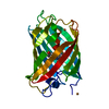
| ||||||||
|---|---|---|---|---|---|---|---|---|---|
| 1 |
| ||||||||
| Unit cell |
|
-Components
| #1: Protein | Mass: 24825.068 Da / Num. of mol.: 1 / Fragment: unp residues 44-258 Mutation: H63Y, H80N, L82I, S100T, N101T, Q104A, L110F, A118P, D119N, M151L, R161Y, F162L, D163K, M165E, L179T, K180G, E182D, P183A, I187R, V196K, I199V, S200K, S202K, H210Y, C213V, S217T, K220R, ...Mutation: H63Y, H80N, L82I, S100T, N101T, Q104A, L110F, A118P, D119N, M151L, R161Y, F162L, D163K, M165E, L179T, K180G, E182D, P183A, I187R, V196K, I199V, S200K, S202K, H210Y, C213V, S217T, K220R, V224A, I239C, Y246H, L251V, N254S, Y259N, L261T, L262F Source method: isolated from a genetically manipulated source Source: (gene. exp.) Clavularia sp. (invertebrata) / Plasmid: pET303 / Production host:  | ||
|---|---|---|---|
| #2: Chemical |   Mass: 63.546 Da / Num. of mol.: 2 / Source method: obtained synthetically / Formula: Cu Mass: 63.546 Da / Num. of mol.: 2 / Source method: obtained synthetically / Formula: Cu#3: Water | ChemComp-HOH / |  Mass: 18.015 Da / Num. of mol.: 310 / Source method: isolated from a natural source / Formula: H2O Mass: 18.015 Da / Num. of mol.: 310 / Source method: isolated from a natural source / Formula: H2O |
-Experimental details
-Experiment
| Experiment | Method: X-RAY DIFFRACTION / Number of used crystals: 1 |
|---|
- Sample preparation
| Crystal | Density Matthews: 2.19 Å3/Da / Density % sol: 43.92 % |
|---|---|
| Crystal grow | Temperature: 293 K / Method: vapor diffusion, sitting drop / pH: 6.5 Details: 25.9% PEG3000, 100mM MES, pH 6.5, VAPOR DIFFUSION, SITTING DROP, temperature 293K |
-Data collection
| Diffraction | Mean temperature: 100 K |
|---|---|
| Diffraction source | Source: ROTATING ANODE / Type: BRUKER AXS MICROSTAR / Wavelength: 1.5418 Å |
| Detector | Type: Bruker Platinum 135 / Detector: CCD / Date: Jun 6, 2014 |
| Radiation | Monochromator: BRUKER MICROSTAR MICRO-FOCUS (MONTEL OPTICS) / Protocol: SINGLE WAVELENGTH / Monochromatic (M) / Laue (L): M / Scattering type: x-ray |
| Radiation wavelength | Wavelength: 1.5418 Å / Relative weight: 1 |
| Reflection | Resolution: 1.55→70 Å / Num. all: 32546 / Num. obs: 32526 / % possible obs: 99.9 % / Observed criterion σ(F): 2 / Observed criterion σ(I): 2 / Rmerge(I) obs: 0.04 / Net I/σ(I): 17 |
| Reflection shell | Resolution: 1.55→1.65 Å / Rmerge(I) obs: 0.39 / Mean I/σ(I) obs: 4.7 / % possible all: 99.8 |
- Processing
| Software |
| ||||||||||||||||||||||||||||||||||||||||||||||||||||||||||||||||||||||||||||||||||||||||||||||||||||||||||||||||||||||||
|---|---|---|---|---|---|---|---|---|---|---|---|---|---|---|---|---|---|---|---|---|---|---|---|---|---|---|---|---|---|---|---|---|---|---|---|---|---|---|---|---|---|---|---|---|---|---|---|---|---|---|---|---|---|---|---|---|---|---|---|---|---|---|---|---|---|---|---|---|---|---|---|---|---|---|---|---|---|---|---|---|---|---|---|---|---|---|---|---|---|---|---|---|---|---|---|---|---|---|---|---|---|---|---|---|---|---|---|---|---|---|---|---|---|---|---|---|---|---|---|---|---|
| Refinement | Method to determine structure: MOLECULAR REPLACEMENT Starting model: pdb entry 4Q9W Resolution: 1.55→10 Å / Cor.coef. Fo:Fc: 0.969 / Cor.coef. Fo:Fc free: 0.955 / SU B: 3.44 / SU ML: 0.054 / Cross valid method: THROUGHOUT / ESU R: 0.099 / ESU R Free: 0.08 / Stereochemistry target values: MAXIMUM LIKELIHOOD / Details: HYDROGENS HAVE BEEN ADDED IN THE RIDING POSITIONS
| ||||||||||||||||||||||||||||||||||||||||||||||||||||||||||||||||||||||||||||||||||||||||||||||||||||||||||||||||||||||||
| Solvent computation | Ion probe radii: 0.8 Å / Shrinkage radii: 0.8 Å / VDW probe radii: 1.2 Å / Solvent model: MASK | ||||||||||||||||||||||||||||||||||||||||||||||||||||||||||||||||||||||||||||||||||||||||||||||||||||||||||||||||||||||||
| Displacement parameters | Biso mean: 14.456 Å2
| ||||||||||||||||||||||||||||||||||||||||||||||||||||||||||||||||||||||||||||||||||||||||||||||||||||||||||||||||||||||||
| Refinement step | Cycle: LAST / Resolution: 1.55→10 Å
| ||||||||||||||||||||||||||||||||||||||||||||||||||||||||||||||||||||||||||||||||||||||||||||||||||||||||||||||||||||||||
| Refine LS restraints |
| ||||||||||||||||||||||||||||||||||||||||||||||||||||||||||||||||||||||||||||||||||||||||||||||||||||||||||||||||||||||||
| LS refinement shell | Resolution: 1.55→1.589 Å / Total num. of bins used: 20
| ||||||||||||||||||||||||||||||||||||||||||||||||||||||||||||||||||||||||||||||||||||||||||||||||||||||||||||||||||||||||
| Refinement TLS params. | Method: refined / Origin x: 15.9045 Å / Origin y: -2.5472 Å / Origin z: -3.564 Å
| ||||||||||||||||||||||||||||||||||||||||||||||||||||||||||||||||||||||||||||||||||||||||||||||||||||||||||||||||||||||||
| Refinement TLS group |
|
