 Movie
Movie Controller


+ Open data

- Basic information
| Entry | Database: PDB / ID: 4r0o | ||||||
|---|---|---|---|---|---|---|---|
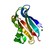
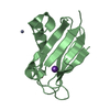
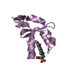
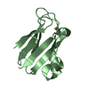
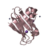
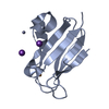
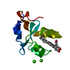
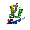
| Title | Crystal structure of PEGylated plastocyanin at 4.2 A resolution | ||||||
 Components Components | Plastocyanin | ||||||
 Keywords Keywords | ELECTRON TRANSPORT / PEGylation | ||||||
| Function / homology |  Function and homology information Function and homology informationplasma membrane-derived thylakoid membrane / electron transfer activity / copper ion binding Similarity search - Function | ||||||
| Biological species |  Phormidium laminosum (bacteria) Phormidium laminosum (bacteria) | ||||||
| Method |  X-RAY DIFFRACTION / SYNCHROTRON / MOLECULAR REPLACEMENT / Resolution: 4.2 Å X-RAY DIFFRACTION / SYNCHROTRON / MOLECULAR REPLACEMENT / Resolution: 4.2 Å | ||||||
 Authors Authors | Cattani, G. / Vogeley, L. / Crowley, P.B. | ||||||
 Citation Citation | Journal: NAT.CHEM. / Year: 2015 Title: Structure of a PEGylated protein reveals a highly porous double-helical assembly. Authors: Cattani, G. / Vogeley, L. / Crowley, P.B. | ||||||
| History |
|
- Structure visualization
| Structure viewer | Molecule: MolmilJmol/JSmol |
|---|
- Downloads & links
-Download
| PDBx/mmCIF format | 4r0o.cif.gz | 93.2 KB | Display | PDBx/mmCIF format |
|---|---|---|---|---|
| PDB format | pdb4r0o.ent.gz | 71.7 KB | Display | PDB format |
| PDBx/mmJSON format | 4r0o.json.gz | Tree view | PDBx/mmJSON format | |
| Others |  Other downloads Other downloads |
-Validation report
| Arichive directory | https://data.pdbj.org/pub/pdb/validation_reports/r0/4r0oftp://data.pdbj.org/pub/pdb/validation_reports/r0/4r0o | HTTPS FTP |
|---|
-Related structure data
| Related structure data |  2w88S S: Starting model for refinement |
|---|---|
| Similar structure data |


-Links
 PDBj
PDBj- Assembly
| Deposited unit | 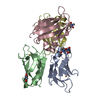
| ||||||||
|---|---|---|---|---|---|---|---|---|---|
| 1 | 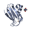
| ||||||||
| 2 | 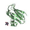
| ||||||||
| 3 | 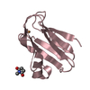
| ||||||||
| 4 | 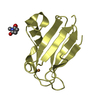
| ||||||||
| Unit cell |
|
-Components
| #1: Protein | Mass: 11551.052 Da / Num. of mol.: 4 / Fragment: UNP residues 35-139 Source method: isolated from a genetically manipulated source Source: (gene. exp.) Phormidium laminosum (bacteria) / Gene: petE / Production host: #2: Chemical | ChemComp-CU /   Mass: 63.546 Da / Num. of mol.: 4 / Source method: obtained synthetically / Formula: Cu Mass: 63.546 Da / Num. of mol.: 4 / Source method: obtained synthetically / Formula: Cu#3: Chemical | ChemComp-LCY /   Mass: 113.115 Da / Num. of mol.: 4 / Source method: obtained synthetically / Formula: C5H7NO2 Mass: 113.115 Da / Num. of mol.: 4 / Source method: obtained synthetically / Formula: C5H7NO2Has protein modification | Y | |
|---|
-Experimental details
-Experiment
| Experiment | Method: X-RAY DIFFRACTION / Number of used crystals: 1 |
|---|
- Sample preparation
| Crystal | Density Matthews: 6.28 Å3/Da / Density % sol: 80.42 % |
|---|---|
| Crystal grow | Temperature: 293.15 K / Method: vapor diffusion, hanging drop / pH: 6.3 Details: 48 % AMMONIUM SULPHATE, 30 mM POTASSIUM FERRICYANIDE, 100 mM SODIUM ACETATE, pH 6.3, VAPOR DIFFUSION, HANGING DROP, temperature 293.15K |
-Data collection
| Diffraction | Mean temperature: 100 K |
|---|---|
| Diffraction source | Source: SYNCHROTRON / Site: Diamond  / Beamline: I24 / Wavelength: 0.9686 Å / Beamline: I24 / Wavelength: 0.9686 Å |
| Detector | Type: PSI PILATUS 6M / Detector: PIXEL / Date: Oct 13, 2013 |
| Radiation | Protocol: SINGLE WAVELENGTH / Monochromatic (M) / Laue (L): M / Scattering type: x-ray |
| Radiation wavelength | Wavelength: 0.9686 Å / Relative weight: 1 |
| Reflection | Resolution: 4.2→44.47 Å / Num. all: 9100 / Num. obs: 9064 / % possible obs: 99.6 % / Redundancy: 5.6 % / Biso Wilson estimate: 174.864 Å2 / Rmerge(I) obs: 0.35 / Rsym value: 0.379 / Net I/σ(I): 5.4 |
| Reflection shell | Resolution: 4.2→4.31 Å / Redundancy: 5.9 % / Rmerge(I) obs: 0.01062 / Mean I/σ(I) obs: 1.7 / Num. unique all: 662 / Rsym value: 0.01152 / % possible all: 100 |
- Processing
| Software |
| ||||||||||||||||||||||||||||
|---|---|---|---|---|---|---|---|---|---|---|---|---|---|---|---|---|---|---|---|---|---|---|---|---|---|---|---|---|---|
| Refinement | Method to determine structure: MOLECULAR REPLACEMENT Starting model: 2w88 Resolution: 4.2→43.035 Å / SU ML: 0.58 / Cross valid method: THROUGHOUT / σ(F): 1.36 / Phase error: 24.86 / Stereochemistry target values: ML
| ||||||||||||||||||||||||||||
| Solvent computation | Shrinkage radii: 0.9 Å / VDW probe radii: 1.11 Å / Solvent model: FLAT BULK SOLVENT MODEL | ||||||||||||||||||||||||||||
| Displacement parameters | Biso mean: 121.577 Å2
| ||||||||||||||||||||||||||||
| Refinement step | Cycle: LAST / Resolution: 4.2→43.035 Å
| ||||||||||||||||||||||||||||
| Refine LS restraints |
| ||||||||||||||||||||||||||||
| LS refinement shell |
|
