 Movie
Movie Controller

[English] 日本語
 Yorodumi
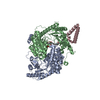
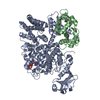
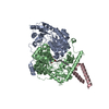
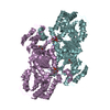
- PDB-4qyr: Streptomyces platensis isomigrastatin ketosynthase domain MgsE KS3 -
+ Open data

- Basic information
| Entry | Database: PDB / ID: 4qyr | ||||||
|---|---|---|---|---|---|---|---|
| Title | Streptomyces platensis isomigrastatin ketosynthase domain MgsE KS3 | ||||||
 Components Components | AT-less polyketide synthase | ||||||
 Keywords Keywords | TRANSFERASE / Structural Genomics / PSI-Biology / Midwest Center for Structural Genomics / MCSG / Enzyme Discovery for Natural Product Biosynthesis / NatPro / alpha-beta structure | ||||||
| Function / homology |  Function and homology information Function and homology informationDIM/DIP cell wall layer assembly / fatty acid synthase activity / phosphopantetheine binding / 3-oxoacyl-[acyl-carrier-protein] synthase activity / antibiotic biosynthetic process / fatty acid biosynthetic process / plasma membrane / cytoplasm Similarity search - Function | ||||||
| Biological species |  Streptomyces platensis subsp. rosaceus (bacteria) Streptomyces platensis subsp. rosaceus (bacteria) | ||||||
| Method |  X-RAY DIFFRACTION / SYNCHROTRON / MOLECULAR REPLACEMENT / Resolution: 2.902 Å X-RAY DIFFRACTION / SYNCHROTRON / MOLECULAR REPLACEMENT / Resolution: 2.902 Å | ||||||
 Authors Authors | Kim, Y. / Li, H. / Endres, M. / Babnigg, J. / Bingman, C.A. / Yennamalli, R. / Lohman, J.R. / Ma, M. / Shen, B. / Phillips Jr., G.N. ...Kim, Y. / Li, H. / Endres, M. / Babnigg, J. / Bingman, C.A. / Yennamalli, R. / Lohman, J.R. / Ma, M. / Shen, B. / Phillips Jr., G.N. / Joachimiak, A. / Midwest Center for Structural Genomics (MCSG) / Enzyme Discovery for Natural Product Biosynthesis (NatPro) | ||||||
 Citation Citation | Journal: Proc.Natl.Acad.Sci.USA / Year: 2015 Title: Structural and evolutionary relationships of "AT-less" type I polyketide synthase ketosynthases. Authors: Lohman, J.R. / Ma, M. / Osipiuk, J. / Nocek, B. / Kim, Y. / Chang, C. / Cuff, M. / Mack, J. / Bigelow, L. / Li, H. / Endres, M. / Babnigg, G. / Joachimiak, A. / Phillips, G.N. / Shen, B. | ||||||
| History |
|
- Structure visualization
| Structure viewer | Molecule: MolmilJmol/JSmol |
|---|
- Downloads & links
-Download
| PDBx/mmCIF format | 4qyr.cif.gz | 243.3 KB | Display | PDBx/mmCIF format |
|---|---|---|---|---|
| PDB format | pdb4qyr.ent.gz | 195.9 KB | Display | PDB format |
| PDBx/mmJSON format | 4qyr.json.gz | Tree view | PDBx/mmJSON format | |
| Others |  Other downloads Other downloads |
-Validation report
| Arichive directory | https://data.pdbj.org/pub/pdb/validation_reports/qy/4qyrftp://data.pdbj.org/pub/pdb/validation_reports/qy/4qyr | HTTPS FTP |
|---|
-Related structure data
| Related structure data |  4opeC  4opfC  4oqjC 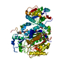 4tktC  4wkyC 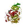 4zdnC 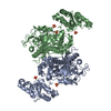 4na1S C: citing same article ( S: Starting model for refinement |
|---|---|
| Similar structure data | |
| Other databases |


-Links
 PDBj
PDBj


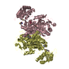
- Assembly
| Deposited unit | 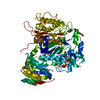
| ||||||||
|---|---|---|---|---|---|---|---|---|---|
| 1 | 
| ||||||||
| Unit cell |
| ||||||||
| Details | a dimer is generated by applying x,y,z and y,x,-z to the asymmetric unit. |
-Components
| #1: Protein | Mass: 65851.141 Da / Num. of mol.: 1 / Fragment: UNP residues 2542-3153 Source method: isolated from a genetically manipulated source Source: (gene. exp.) Streptomyces platensis subsp. rosaceus (bacteria)Plasmid: pMCSG73 / Production host: |
|---|---|
| #2: Chemical | ChemComp-GOL /   Mass: 92.094 Da / Num. of mol.: 1 / Source method: obtained synthetically / Formula: C3H8O3 Mass: 92.094 Da / Num. of mol.: 1 / Source method: obtained synthetically / Formula: C3H8O3 |
| #3: Chemical | ChemComp-ACY /   Mass: 60.052 Da / Num. of mol.: 1 / Source method: obtained synthetically / Formula: C2H4O2 Mass: 60.052 Da / Num. of mol.: 1 / Source method: obtained synthetically / Formula: C2H4O2 |
| #4: Chemical | ChemComp-CL /   Mass: 35.453 Da / Num. of mol.: 1 / Source method: obtained synthetically / Formula: Cl Mass: 35.453 Da / Num. of mol.: 1 / Source method: obtained synthetically / Formula: Cl |
| #5: Water | ChemComp-HOH /  Mass: 18.015 Da / Num. of mol.: 45 / Source method: isolated from a natural source / Formula: H2O Mass: 18.015 Da / Num. of mol.: 45 / Source method: isolated from a natural source / Formula: H2O |
| Has protein modification | Y |
-Experimental details
-Experiment
| Experiment | Method: X-RAY DIFFRACTION / Number of used crystals: 1 |
|---|
- Sample preparation
| Crystal | Density Matthews: 3.14 Å3/Da / Density % sol: 60.81 % |
|---|---|
| Crystal grow | Temperature: 289 K / Method: vapor diffusion, sitting drop / pH: 7.5 Details: 0.1 M sodium acetate, 0.1 M HEPES pH 7.5, 22%(w/v) PEG4000, VAPOR DIFFUSION, SITTING DROP, temperature 289K |
-Data collection
| Diffraction | Mean temperature: 100 K |
|---|---|
| Diffraction source | Source: SYNCHROTRON / Site: APS  / Beamline: 19-ID / Wavelength: 0.97924 Å / Beamline: 19-ID / Wavelength: 0.97924 Å |
| Detector | Type: ADSC QUANTUM 315r / Detector: CCD / Date: Mar 17, 2014 / Details: mirrors |
| Radiation | Monochromator: double crystal monochromator / Protocol: SINGLE WAVELENGTH / Monochromatic (M) / Laue (L): M / Scattering type: x-ray |
| Radiation wavelength | Wavelength: 0.97924 Å / Relative weight: 1 |
| Reflection | Resolution: 2.9→50 Å / Num. all: 18196 / Num. obs: 18196 / % possible obs: 98.2 % / Observed criterion σ(F): 0 / Observed criterion σ(I): 0 / Redundancy: 4.1 % / Biso Wilson estimate: 53.25 Å2 / Rsym value: 0.103 / Net I/σ(I): 11.1 |
| Reflection shell | Resolution: 2.9→2.95 Å / Redundancy: 4 % / Mean I/σ(I) obs: 2.3 / Num. unique all: 888 / Rsym value: 0.754 / % possible all: 96.9 |
- Processing
| Software |
| |||||||||||||||||||||||||||||||||||||||||||||||||||||||||||||||||||||||||||||||||||||||||||||||||||||||||||||||||||||||||||||||||||||||||||||||||||||||||||||||||||||||||||||||
|---|---|---|---|---|---|---|---|---|---|---|---|---|---|---|---|---|---|---|---|---|---|---|---|---|---|---|---|---|---|---|---|---|---|---|---|---|---|---|---|---|---|---|---|---|---|---|---|---|---|---|---|---|---|---|---|---|---|---|---|---|---|---|---|---|---|---|---|---|---|---|---|---|---|---|---|---|---|---|---|---|---|---|---|---|---|---|---|---|---|---|---|---|---|---|---|---|---|---|---|---|---|---|---|---|---|---|---|---|---|---|---|---|---|---|---|---|---|---|---|---|---|---|---|---|---|---|---|---|---|---|---|---|---|---|---|---|---|---|---|---|---|---|---|---|---|---|---|---|---|---|---|---|---|---|---|---|---|---|---|---|---|---|---|---|---|---|---|---|---|---|---|---|---|---|---|---|
| Refinement | Method to determine structure: MOLECULAR REPLACEMENT Starting model: PDB entry 4NA1 Resolution: 2.902→36.354 Å / SU ML: 0.31 / Isotropic thermal model: mixed / Cross valid method: THROUGHOUT / σ(F): 0 / Phase error: 20.59 / Stereochemistry target values: ML
| |||||||||||||||||||||||||||||||||||||||||||||||||||||||||||||||||||||||||||||||||||||||||||||||||||||||||||||||||||||||||||||||||||||||||||||||||||||||||||||||||||||||||||||||
| Solvent computation | Shrinkage radii: 0.9 Å / VDW probe radii: 1.11 Å / Solvent model: FLAT BULK SOLVENT MODEL | |||||||||||||||||||||||||||||||||||||||||||||||||||||||||||||||||||||||||||||||||||||||||||||||||||||||||||||||||||||||||||||||||||||||||||||||||||||||||||||||||||||||||||||||
| Displacement parameters | Biso mean: 59.4 Å2 | |||||||||||||||||||||||||||||||||||||||||||||||||||||||||||||||||||||||||||||||||||||||||||||||||||||||||||||||||||||||||||||||||||||||||||||||||||||||||||||||||||||||||||||||
| Refinement step | Cycle: LAST / Resolution: 2.902→36.354 Å
| |||||||||||||||||||||||||||||||||||||||||||||||||||||||||||||||||||||||||||||||||||||||||||||||||||||||||||||||||||||||||||||||||||||||||||||||||||||||||||||||||||||||||||||||
| Refine LS restraints |
| |||||||||||||||||||||||||||||||||||||||||||||||||||||||||||||||||||||||||||||||||||||||||||||||||||||||||||||||||||||||||||||||||||||||||||||||||||||||||||||||||||||||||||||||
| LS refinement shell | Refine-ID: X-RAY DIFFRACTION
| |||||||||||||||||||||||||||||||||||||||||||||||||||||||||||||||||||||||||||||||||||||||||||||||||||||||||||||||||||||||||||||||||||||||||||||||||||||||||||||||||||||||||||||||
| Refinement TLS params. | Method: refined / Refine-ID: X-RAY DIFFRACTION
| |||||||||||||||||||||||||||||||||||||||||||||||||||||||||||||||||||||||||||||||||||||||||||||||||||||||||||||||||||||||||||||||||||||||||||||||||||||||||||||||||||||||||||||||
| Refinement TLS group |
|
