 Movie
Movie Controller

[English] 日本語
 Yorodumi
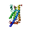
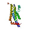
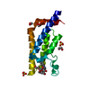
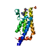
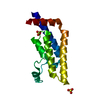
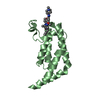
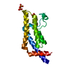
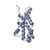
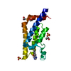
- PDB-4qsw: Structure of the bromodomain of human ATPase family AAA domain-co... -
+ Open data

- Basic information
| Entry | Database: PDB / ID: 4qsw | ||||||
|---|---|---|---|---|---|---|---|
| Title | Structure of the bromodomain of human ATPase family AAA domain-containing protein 2 (ATAD2) in complex with 5-methyl uridine | ||||||
 Components Components | ATPase family AAA domain-containing protein 2 | ||||||
 Keywords Keywords | SIGNALING PROTEIN / Structural Genomics Consortium (SGC) / bromodomain / actyl-lysine binding / ATPase family AAA domain-containing protein 2 / epigenetics | ||||||
| Function / homology |  Function and homology information Function and homology informationnucleosome disassembly / Hydrolases; Acting on acid anhydrides; In phosphorus-containing anhydrides / TFAP2 (AP-2) family regulates transcription of growth factors and their receptors / transcription initiation-coupled chromatin remodeling / nucleosome assembly / histone binding / chromatin binding / positive regulation of DNA-templated transcription / ATP hydrolysis activity / extracellular exosome ...nucleosome disassembly / Hydrolases; Acting on acid anhydrides; In phosphorus-containing anhydrides / TFAP2 (AP-2) family regulates transcription of growth factors and their receptors / transcription initiation-coupled chromatin remodeling / nucleosome assembly / histone binding / chromatin binding / positive regulation of DNA-templated transcription / ATP hydrolysis activity / extracellular exosome / nucleoplasm / ATP binding / nucleus Similarity search - Function | ||||||
| Biological species |  Homo sapiens (human) Homo sapiens (human) | ||||||
| Method |  X-RAY DIFFRACTION / MOLECULAR REPLACEMENT / Resolution: 1.8 Å X-RAY DIFFRACTION / MOLECULAR REPLACEMENT / Resolution: 1.8 Å | ||||||
 Authors Authors | Chaikuad, A. / Felletar, I. / von Delft, F. / Arrowsmith, C.H. / Edwards, A.M. / Bountra, C. / Knapp, S. / Structural Genomics Consortium (SGC) | ||||||
 Citation Citation | Journal: MedChemComm / Year: 2014 Title: Structure-based approaches towards identification of fragments for the low-druggability ATAD2 bromodomain Authors: Chaikuad, A. / Petros, A.M. / Fedorov, O. / Xu, J. / Knapp, S. | ||||||
| History |
|
- Structure visualization
| Structure viewer | Molecule: MolmilJmol/JSmol |
|---|
- Downloads & links
-Download
| PDBx/mmCIF format | 4qsw.cif.gz | 79.5 KB | Display | PDBx/mmCIF format |
|---|---|---|---|---|
| PDB format | pdb4qsw.ent.gz | 58.9 KB | Display | PDB format |
| PDBx/mmJSON format | 4qsw.json.gz | Tree view | PDBx/mmJSON format | |
| Others |  Other downloads Other downloads |
-Validation report
| Arichive directory | https://data.pdbj.org/pub/pdb/validation_reports/qs/4qswftp://data.pdbj.org/pub/pdb/validation_reports/qs/4qsw | HTTPS FTP |
|---|
-Related structure data
| Related structure data | 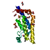 4qspC 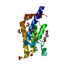 4qsqC 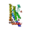 4qsrC 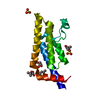 4qssC 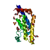 4qstC 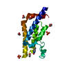 4qsuC  4qsvC 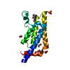 4qsxC  3daiS C: citing same article ( S: Starting model for refinement |
|---|---|
| Similar structure data |

-Links
 PDBj
PDBj


- Assembly
| Deposited unit | 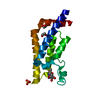
| ||||||||
|---|---|---|---|---|---|---|---|---|---|
| 1 |
| ||||||||
| Unit cell |
| ||||||||
| Components on special symmetry positions |
|
-Components
| #1: Protein | Mass: 15453.514 Da / Num. of mol.: 1 / Fragment: bromodomain (residues 981-1108) Source method: isolated from a genetically manipulated source Source: (gene. exp.) Homo sapiens (human) / Gene: ATAD2, L16, PRO2000 / Plasmid: pNIC28-Bsa4 / Production host:  | ||||
|---|---|---|---|---|---|
| #2: Chemical | ChemComp-SO4 /   Mass: 96.063 Da / Num. of mol.: 1 / Source method: obtained synthetically / Formula: SO4 Mass: 96.063 Da / Num. of mol.: 1 / Source method: obtained synthetically / Formula: SO4 | ||||
| #3: Chemical | ChemComp-EDO /   Mass: 62.068 Da / Num. of mol.: 5 / Source method: obtained synthetically / Formula: C2H6O2 Mass: 62.068 Da / Num. of mol.: 5 / Source method: obtained synthetically / Formula: C2H6O2#4: Chemical | ChemComp-38T / |   Mass: 258.228 Da / Num. of mol.: 1 / Source method: obtained synthetically / Formula: C10H14N2O6 Mass: 258.228 Da / Num. of mol.: 1 / Source method: obtained synthetically / Formula: C10H14N2O6#5: Water | ChemComp-HOH / |  Mass: 18.015 Da / Num. of mol.: 243 / Source method: isolated from a natural source / Formula: H2O Mass: 18.015 Da / Num. of mol.: 243 / Source method: isolated from a natural source / Formula: H2O |
-Experimental details
-Experiment
| Experiment | Method: X-RAY DIFFRACTION / Number of used crystals: 1 |
|---|
- Sample preparation
| Crystal | Density Matthews: 4.09 Å3/Da / Density % sol: 69.96 % |
|---|---|
| Crystal grow | Temperature: 277.15 K / Method: vapor diffusion, sitting drop / pH: 5.5 Details: apo crystals grew in 1.8-2.2 M ammonium sulphate, 0.1 M Bis-Tris, pH 5.5-6.5. Soaking performed in 28-32% PEG 3350, 50 mM bis-tris pH 5.5, 50 mM ammonium phosphate and 20% ethylene glycol, ...Details: apo crystals grew in 1.8-2.2 M ammonium sulphate, 0.1 M Bis-Tris, pH 5.5-6.5. Soaking performed in 28-32% PEG 3350, 50 mM bis-tris pH 5.5, 50 mM ammonium phosphate and 20% ethylene glycol, VAPOR DIFFUSION, SITTING DROP, temperature 277.15K |
-Data collection
| Diffraction | Mean temperature: 100 K |
|---|---|
| Diffraction source | Source: ROTATING ANODE / Type: RIGAKU FR-E SUPERBRIGHT / Wavelength: 1.5418 Å |
| Detector | Type: RIGAKU RAXIS IV / Detector: IMAGE PLATE / Date: May 10, 2013 |
| Radiation | Monochromator: Flat graphite crystal / Protocol: SINGLE WAVELENGTH / Monochromatic (M) / Laue (L): M / Scattering type: x-ray |
| Radiation wavelength | Wavelength: 1.5418 Å / Relative weight: 1 |
| Reflection | Resolution: 1.8→34.59 Å / Num. all: 24186 / Num. obs: 24161 / % possible obs: 98.3 % / Observed criterion σ(F): 0 / Observed criterion σ(I): 0 / Redundancy: 9.3 % / Biso Wilson estimate: 27.8 Å2 / Rmerge(I) obs: 0.08 / Net I/σ(I): 16.2 |
| Reflection shell | Resolution: 1.8→1.9 Å / Redundancy: 9.4 % / Rmerge(I) obs: 0.798 / Mean I/σ(I) obs: 2.6 / Num. unique all: 3381 / % possible all: 96.5 |
- Processing
| Software |
| |||||||||||||||||||||||||||||||||||||||||||||||||||||||||||||||||||||||||||||||||||||||||||||||||||||||||
|---|---|---|---|---|---|---|---|---|---|---|---|---|---|---|---|---|---|---|---|---|---|---|---|---|---|---|---|---|---|---|---|---|---|---|---|---|---|---|---|---|---|---|---|---|---|---|---|---|---|---|---|---|---|---|---|---|---|---|---|---|---|---|---|---|---|---|---|---|---|---|---|---|---|---|---|---|---|---|---|---|---|---|---|---|---|---|---|---|---|---|---|---|---|---|---|---|---|---|---|---|---|---|---|---|---|---|
| Refinement | Method to determine structure: MOLECULAR REPLACEMENT Starting model: pdb id: 3DAI Resolution: 1.8→30.93 Å / Cor.coef. Fo:Fc: 0.966 / Cor.coef. Fo:Fc free: 0.95 / SU B: 3.32 / SU ML: 0.055 / Cross valid method: THROUGHOUT / σ(F): 0 / σ(I): 2 / ESU R: 0.09 / ESU R Free: 0.095 / Stereochemistry target values: MAXIMUM LIKELIHOOD / Details: HYDROGENS HAVE BEEN ADDED IN THE RIDING POSITIONS
| |||||||||||||||||||||||||||||||||||||||||||||||||||||||||||||||||||||||||||||||||||||||||||||||||||||||||
| Solvent computation | Ion probe radii: 0.8 Å / Shrinkage radii: 0.8 Å / VDW probe radii: 1.2 Å / Solvent model: MASK | |||||||||||||||||||||||||||||||||||||||||||||||||||||||||||||||||||||||||||||||||||||||||||||||||||||||||
| Displacement parameters | Biso mean: 31.416 Å2
| |||||||||||||||||||||||||||||||||||||||||||||||||||||||||||||||||||||||||||||||||||||||||||||||||||||||||
| Refine analyze | Luzzati coordinate error obs: 0.21 Å | |||||||||||||||||||||||||||||||||||||||||||||||||||||||||||||||||||||||||||||||||||||||||||||||||||||||||
| Refinement step | Cycle: LAST / Resolution: 1.8→30.93 Å
| |||||||||||||||||||||||||||||||||||||||||||||||||||||||||||||||||||||||||||||||||||||||||||||||||||||||||
| Refine LS restraints |
| |||||||||||||||||||||||||||||||||||||||||||||||||||||||||||||||||||||||||||||||||||||||||||||||||||||||||
| LS refinement shell | Resolution: 1.801→1.848 Å / Total num. of bins used: 20
| |||||||||||||||||||||||||||||||||||||||||||||||||||||||||||||||||||||||||||||||||||||||||||||||||||||||||
| Refinement TLS params. | Method: refined / Refine-ID: X-RAY DIFFRACTION
| |||||||||||||||||||||||||||||||||||||||||||||||||||||||||||||||||||||||||||||||||||||||||||||||||||||||||
| Refinement TLS group |
|
