 Movie
Movie Controller


+ Open data

- Basic information
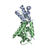
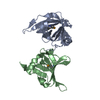
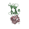
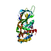
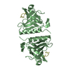
| Entry | Database: PDB / ID: 4qki | ||||||
|---|---|---|---|---|---|---|---|
| Title | Dimeric form of human LLT1, a ligand for NKR-P1 | ||||||
 Components Components | C-type lectin domain family 2 member D | ||||||
 Keywords Keywords | IMMUNE SYSTEM / C-type lectin like fold / ligand for human receptor NKR-P1 / glycosylation / deglycosylated after the first GlcNac unit / anchored in membrane on cell surface | ||||||
| Function / homology |  Function and homology information Function and homology informationImmunoregulatory interactions between a Lymphoid and a non-Lymphoid cell / transmembrane signaling receptor activity / carbohydrate binding / cell surface receptor signaling pathway / external side of plasma membrane / cell surface / endoplasmic reticulum / membrane / plasma membrane Similarity search - Function | ||||||
| Biological species |  Homo sapiens (human) Homo sapiens (human) | ||||||
| Method |  X-RAY DIFFRACTION / SYNCHROTRON / MOLECULAR REPLACEMENT / Resolution: 1.8 Å X-RAY DIFFRACTION / SYNCHROTRON / MOLECULAR REPLACEMENT / Resolution: 1.8 Å | ||||||
 Authors Authors | Skalova, T. / Blaha, J. / Harlos, K. / Duskova, J. / Koval, T. / Stransky, J. / Hasek, J. / Vanek, O. / Dohnalek, J. | ||||||
 Citation Citation | Journal: Acta Crystallogr.,Sect.D / Year: 2015 Title: Four crystal structures of human LLT1, a ligand of human NKR-P1, in varied glycosylation and oligomerization states Authors: Skalova, T. / Blaha, J. / Harlos, K. / Duskova, J. / Koval, T. / Stransky, J. / Hasek, J. / Vanek, O. / Dohnalek, J. | ||||||
| History |
|
- Structure visualization
| Structure viewer | Molecule: MolmilJmol/JSmol |
|---|
- Downloads & links
-Download
| PDBx/mmCIF format | 4qki.cif.gz | 69.1 KB | Display | PDBx/mmCIF format |
|---|---|---|---|---|
| PDB format | pdb4qki.ent.gz | 51.1 KB | Display | PDB format |
| PDBx/mmJSON format | 4qki.json.gz | Tree view | PDBx/mmJSON format | |
| Others |  Other downloads Other downloads |
-Validation report
| Arichive directory | https://data.pdbj.org/pub/pdb/validation_reports/qk/4qkiftp://data.pdbj.org/pub/pdb/validation_reports/qk/4qki | HTTPS FTP |
|---|
-Related structure data
| Related structure data | 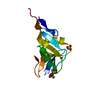 4qkgC  4qkhC 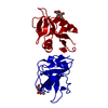 4qkjC 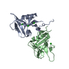 3hupS C: citing same article ( S: Starting model for refinement |
|---|---|
| Similar structure data |


-Links
 PDBj
PDBj


- Assembly
| Deposited unit | 
| ||||||||
|---|---|---|---|---|---|---|---|---|---|
| 1 |
| ||||||||
| 2 | 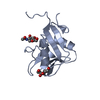
| ||||||||
| 3 | 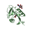
| ||||||||
| Unit cell |
|
-Components
| #1: Protein | Mass: 15692.363 Da / Num. of mol.: 2 / Fragment: extracellular part, UNP residues 72-191 / Mutation: H176C Source method: isolated from a genetically manipulated source Details: protein secreted into medium / Source: (gene. exp.) Homo sapiens (human) / Gene: CLAX, CLEC2B, CLEC2D, LLT1, OCIL / Plasmid: pTT28 / Cell line (production host): HEK293S GnTI- / Production host: HOMO SAPIENS (human) / References: UniProt: Q9UHP7#2: Sugar | ChemComp-NAG /   Type: D-saccharide, beta linking / Mass: 221.208 Da / Num. of mol.: 4 Type: D-saccharide, beta linking / Mass: 221.208 Da / Num. of mol.: 4Source method: isolated from a genetically manipulated source Formula: C8H15NO6 #3: Water | ChemComp-HOH / |  Mass: 18.015 Da / Num. of mol.: 167 / Source method: isolated from a natural source / Formula: H2O Mass: 18.015 Da / Num. of mol.: 167 / Source method: isolated from a natural source / Formula: H2OHas protein modification | Y | |
|---|
-Experimental details
-Experiment
| Experiment | Method: X-RAY DIFFRACTION / Number of used crystals: 1 |
|---|
- Sample preparation
| Crystal | Density Matthews: 1.64 Å3/Da / Density % sol: 25 % Description: THE ENTRY CONTAINS FRIEDEL PAIRS IN F_PLUS/MINUS COLUMNS. |
|---|---|
| Crystal grow | Temperature: 294 K / Method: vapor diffusion, sitting drop / pH: 7 Details: 30%(w/v) polyethylene glycol 6000, 0.1M HEPES, pH 7.0, VAPOR DIFFUSION, SITTING DROP, temperature 294K |
-Data collection
| Diffraction | Mean temperature: 100 K |
|---|---|
| Diffraction source | Source: SYNCHROTRON / Site: Diamond  / Beamline: I02 / Wavelength: 0.92 Å / Beamline: I02 / Wavelength: 0.92 Å |
| Detector | Type: DECTRIS PILATUS 2M / Detector: PIXEL / Date: Jun 30, 2012 / Details: Kirkpatrick Baez mirror pair |
| Radiation | Monochromator: Si(111) double crystal / Protocol: SINGLE WAVELENGTH / Monochromatic (M) / Laue (L): M / Scattering type: x-ray |
| Radiation wavelength | Wavelength: 0.92 Å / Relative weight: 1 |
| Reflection | Resolution: 1.8→43.73 Å / Num. all: 18996 / Num. obs: 18996 / % possible obs: 96.7 % / Observed criterion σ(F): 0 / Observed criterion σ(I): -3.7 / Redundancy: 6 % / Biso Wilson estimate: 17.7 Å2 / Rmerge(I) obs: 0.082 / Net I/σ(I): 11.8 |
| Reflection shell | Resolution: 1.8→1.84 Å / Redundancy: 6.2 % / Rmerge(I) obs: 0.404 / Mean I/σ(I) obs: 3.7 / Num. unique all: 1050 / % possible all: 91.6 |
- Processing
| Software |
| ||||||||||||||||||||||||||||||||||||||||||||||||||||||||||||||||||||||||||||||||||||||||||||||||||||||||||||||||||||||||||||||||||||||||||||||||||||||||||||||||||||||||||||||||||||||
|---|---|---|---|---|---|---|---|---|---|---|---|---|---|---|---|---|---|---|---|---|---|---|---|---|---|---|---|---|---|---|---|---|---|---|---|---|---|---|---|---|---|---|---|---|---|---|---|---|---|---|---|---|---|---|---|---|---|---|---|---|---|---|---|---|---|---|---|---|---|---|---|---|---|---|---|---|---|---|---|---|---|---|---|---|---|---|---|---|---|---|---|---|---|---|---|---|---|---|---|---|---|---|---|---|---|---|---|---|---|---|---|---|---|---|---|---|---|---|---|---|---|---|---|---|---|---|---|---|---|---|---|---|---|---|---|---|---|---|---|---|---|---|---|---|---|---|---|---|---|---|---|---|---|---|---|---|---|---|---|---|---|---|---|---|---|---|---|---|---|---|---|---|---|---|---|---|---|---|---|---|---|---|---|
| Refinement | Method to determine structure: MOLECULAR REPLACEMENT Starting model: 3HUP Resolution: 1.8→43.73 Å / Cor.coef. Fo:Fc: 0.954 / SU B: 2.461 / SU ML: 0.076 / Cross valid method: throughout (except for the last cycle) / σ(F): 0 / ESU R: 0.154 Stereochemistry target values: CCP4 STEREOCHEMISTRY LIBRARY, VERSION 6.4.0 Details: HYDROGENS HAVE BEEN ADDED IN THE RIDING POSITIONS, OTHER REFINEMENT REMARKS: SF FILE CONTAINS FRIEDEL PAIRS UNDER I/F_MINUS AND I/F_PLUS COLUMNS.
| ||||||||||||||||||||||||||||||||||||||||||||||||||||||||||||||||||||||||||||||||||||||||||||||||||||||||||||||||||||||||||||||||||||||||||||||||||||||||||||||||||||||||||||||||||||||
| Solvent computation | Ion probe radii: 0.8 Å / Shrinkage radii: 0.8 Å / VDW probe radii: 1.2 Å / Solvent model: MASK | ||||||||||||||||||||||||||||||||||||||||||||||||||||||||||||||||||||||||||||||||||||||||||||||||||||||||||||||||||||||||||||||||||||||||||||||||||||||||||||||||||||||||||||||||||||||
| Displacement parameters | Biso mean: 21.046 Å2
| ||||||||||||||||||||||||||||||||||||||||||||||||||||||||||||||||||||||||||||||||||||||||||||||||||||||||||||||||||||||||||||||||||||||||||||||||||||||||||||||||||||||||||||||||||||||
| Refinement step | Cycle: LAST / Resolution: 1.8→43.73 Å
| ||||||||||||||||||||||||||||||||||||||||||||||||||||||||||||||||||||||||||||||||||||||||||||||||||||||||||||||||||||||||||||||||||||||||||||||||||||||||||||||||||||||||||||||||||||||
| Refine LS restraints |
|