 Movie
Movie Controller

[English] 日本語
 Yorodumi
- PDB-4qcc: Structure of a cube-shaped, highly porous protein cage designed b... -
+ Open data

- Basic information
| Entry | Database: PDB / ID: 4qcc | ||||||
|---|---|---|---|---|---|---|---|
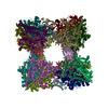
| Title | Structure of a cube-shaped, highly porous protein cage designed by fusing symmetric oligomeric domains | ||||||
 Components Components | 2-dehydro-3-deoxy-6-phosphogalactonate aldolase, peptidyl-prolyl cis-trans isomerase chimera | ||||||
 Keywords Keywords | STRUCTURAL PROTEIN / LYASE / protein design / bionanotechnology / self-assembly / symmetry / porous biomaterials | ||||||
| Function / homology | 2-dehydro-3-deoxy-6-phosphogalactonate aldolase / D-galactonate catabolic process / 2-dehydro-3-deoxy-6-phosphogalactonate aldolase activity / KDPG/KHG aldolase / KDPG and KHG aldolase / Aldolase-type TIM barrel / 2-dehydro-3-deoxy-6-phosphogalactonate aldolase / :  Function and homology information Function and homology information | ||||||
| Biological species |  | ||||||
| Method |  X-RAY DIFFRACTION / MOLECULAR REPLACEMENT / molecular replacement / Resolution: 7.078 Å X-RAY DIFFRACTION / MOLECULAR REPLACEMENT / molecular replacement / Resolution: 7.078 Å | ||||||
 Authors Authors | Lai, Y.-T. / Yeates, T.O. | ||||||
 Citation Citation | Journal: Nat Chem / Year: 2014 Title: Structure of a designed protein cage that self-assembles into a highly porous cube. Authors: Lai, Y.T. / Reading, E. / Hura, G.L. / Tsai, K.L. / Laganowsky, A. / Asturias, F.J. / Tainer, J.A. / Robinson, C.V. / Yeates, T.O. | ||||||
| History |
|
- Structure visualization
| Structure viewer | Molecule: MolmilJmol/JSmol |
|---|
- Downloads & links
-Download
| PDBx/mmCIF format | 4qcc.cif.gz | 111.6 KB | Display | PDBx/mmCIF format |
|---|---|---|---|---|
| PDB format | pdb4qcc.ent.gz | 87.5 KB | Display | PDB format |
| PDBx/mmJSON format | 4qcc.json.gz | Tree view | PDBx/mmJSON format | |
| Others |  Other downloads Other downloads |
-Validation report
| Arichive directory | https://data.pdbj.org/pub/pdb/validation_reports/qc/4qccftp://data.pdbj.org/pub/pdb/validation_reports/qc/4qcc | HTTPS FTP |
|---|
-Related structure data
| Similar structure data |
|---|


-Links
 PDBj
PDBj- Assembly
| Deposited unit | 
| ||||||||||||||||||
|---|---|---|---|---|---|---|---|---|---|---|---|---|---|---|---|---|---|---|---|
| 1 | x 12
| ||||||||||||||||||
| Unit cell |
| ||||||||||||||||||
| Noncrystallographic symmetry (NCS) | NCS domain:
NCS domain segments: Component-ID: _ / Ens-ID: 1 / Beg auth comp-ID: THR / Beg label comp-ID: THR / End auth comp-ID: ALA / End label comp-ID: ALA / Refine code: _ / Auth seq-ID: 5 - 274 / Label seq-ID: 5 - 274
|
-Components
| #1: Protein | Mass: 31233.459 Da / Num. of mol.: 2 / Fragment: SEE REMARK 999 Source method: isolated from a genetically manipulated source Source: (gene. exp.) References: UniProt: Q6BF16, UniProt: U6NBA4, 2-dehydro-3-deoxy-6-phosphogalactonate aldolase Sequence details | PROTEIN IS A DESIGNED CHIMERA COMPRISING RESIDUES 1-203 OF UNP Q6BF16 AND RESIDUES 45-116 OF UNP ...PROTEIN IS A DESIGNED CHIMERA COMPRISING | |
|---|
-Experimental details
-Experiment
| Experiment | Method: X-RAY DIFFRACTION / Number of used crystals: 1 |
|---|
- Sample preparation
| Crystal | Density Matthews: 6.76 Å3/Da / Density % sol: 81.81 % |
|---|---|
| Crystal grow | Temperature: 298 K / Method: vapor diffusion, hanging drop / pH: 6 Details: 0.1 M MES, pH 6.0, 0.6 M ammonium sulfate, 1% PEG3350, VAPOR DIFFUSION, HANGING DROP, temperature 298K |
-Data collection
| Diffraction | Mean temperature: 100 K | |||||||||||||||||||||||||||||||||||||||||||||||||||||||||||||||||||||||||||||||||||||||||||||||||||||||||||||||||||||||||||||||||||||||||||||||||||
|---|---|---|---|---|---|---|---|---|---|---|---|---|---|---|---|---|---|---|---|---|---|---|---|---|---|---|---|---|---|---|---|---|---|---|---|---|---|---|---|---|---|---|---|---|---|---|---|---|---|---|---|---|---|---|---|---|---|---|---|---|---|---|---|---|---|---|---|---|---|---|---|---|---|---|---|---|---|---|---|---|---|---|---|---|---|---|---|---|---|---|---|---|---|---|---|---|---|---|---|---|---|---|---|---|---|---|---|---|---|---|---|---|---|---|---|---|---|---|---|---|---|---|---|---|---|---|---|---|---|---|---|---|---|---|---|---|---|---|---|---|---|---|---|---|---|---|---|---|
| Diffraction source | Source: ROTATING ANODE / Type: RIGAKU / Wavelength: 1.542 Å | |||||||||||||||||||||||||||||||||||||||||||||||||||||||||||||||||||||||||||||||||||||||||||||||||||||||||||||||||||||||||||||||||||||||||||||||||||
| Detector | Type: RIGAKU RAXIS HTC / Detector: IMAGE PLATE / Date: Mar 5, 2014 | |||||||||||||||||||||||||||||||||||||||||||||||||||||||||||||||||||||||||||||||||||||||||||||||||||||||||||||||||||||||||||||||||||||||||||||||||||
| Radiation | Monochromator: Varimax HR / Protocol: SINGLE WAVELENGTH / Monochromatic (M) / Laue (L): M / Scattering type: x-ray | |||||||||||||||||||||||||||||||||||||||||||||||||||||||||||||||||||||||||||||||||||||||||||||||||||||||||||||||||||||||||||||||||||||||||||||||||||
| Radiation wavelength | Wavelength: 1.542 Å / Relative weight: 1 | |||||||||||||||||||||||||||||||||||||||||||||||||||||||||||||||||||||||||||||||||||||||||||||||||||||||||||||||||||||||||||||||||||||||||||||||||||
| Reflection | Resolution: 7.078→96.407 Å / Num. obs: 2526 / % possible obs: 94.7 % / Observed criterion σ(I): -3 / Biso Wilson estimate: 320.857 Å2 / Rmerge(I) obs: 0.073 / Χ2: 0.894 / Net I/σ(I): 15.62 | |||||||||||||||||||||||||||||||||||||||||||||||||||||||||||||||||||||||||||||||||||||||||||||||||||||||||||||||||||||||||||||||||||||||||||||||||||
| Reflection shell | Diffraction-ID: 1
|
-Phasing
| Phasing | Method: molecular replacement | |||||||||
|---|---|---|---|---|---|---|---|---|---|---|
| Phasing MR | Model details: Phaser MODE: MR_AUTO
|
- Processing
| Software |
| |||||||||||||||||||||||||||||||||||||||||||||||||||||||||||||||||||||||||||
|---|---|---|---|---|---|---|---|---|---|---|---|---|---|---|---|---|---|---|---|---|---|---|---|---|---|---|---|---|---|---|---|---|---|---|---|---|---|---|---|---|---|---|---|---|---|---|---|---|---|---|---|---|---|---|---|---|---|---|---|---|---|---|---|---|---|---|---|---|---|---|---|---|---|---|---|---|
| Refinement | Method to determine structure: MOLECULAR REPLACEMENT / Resolution: 7.078→96.407 Å / Cor.coef. Fo:Fc: 0.851 / Cor.coef. Fo:Fc free: 0.769 / WRfactor Rfree: 0.2692 / WRfactor Rwork: 0.258 / FOM work R set: 0.7259 / SU B: 307.178 / SU ML: 2.408 / SU Rfree: 2.983 / Cross valid method: THROUGHOUT / σ(F): 0 / ESU R Free: 2.983 / Stereochemistry target values: MAXIMUM LIKELIHOOD / Details: HYDROGENS HAVE BEEN ADDED IN THE RIDING POSITIONS
| |||||||||||||||||||||||||||||||||||||||||||||||||||||||||||||||||||||||||||
| Solvent computation | Ion probe radii: 0.8 Å / Shrinkage radii: 0.8 Å / VDW probe radii: 1.2 Å / Solvent model: MASK | |||||||||||||||||||||||||||||||||||||||||||||||||||||||||||||||||||||||||||
| Displacement parameters | Biso max: 459.21 Å2 / Biso mean: 289.798 Å2 / Biso min: 100 Å2
| |||||||||||||||||||||||||||||||||||||||||||||||||||||||||||||||||||||||||||
| Refinement step | Cycle: LAST / Resolution: 7.078→96.407 Å
| |||||||||||||||||||||||||||||||||||||||||||||||||||||||||||||||||||||||||||
| Refine LS restraints |
| |||||||||||||||||||||||||||||||||||||||||||||||||||||||||||||||||||||||||||
| Refine LS restraints NCS | Ens-ID: 1 / Number: 15679 / Refine-ID: X-RAY DIFFRACTION / Type: interatomic distance / Rms dev position: 0 Å / Weight position: 0.05
| |||||||||||||||||||||||||||||||||||||||||||||||||||||||||||||||||||||||||||
| LS refinement shell | Resolution: 7.078→7.261 Å / Total num. of bins used: 20
|
