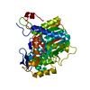
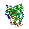
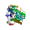
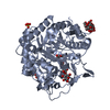
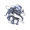
- PDB-4q9t: Crystal structure of Vanderwaltozyma polyspora Nup133 Beta-propel... -
+
Open data
ID or keywords:
Loading...
-
Basic information
Entry
Database: PDB / ID: 4q9t
Title
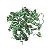
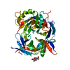
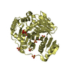
Crystal structure of Vanderwaltozyma polyspora Nup133 Beta-propeller domain
Components
Nucleoporin NUP133
Keywords
PROTEIN TRANSPORT / Nuclear Pore Complex / Nucleoporin / Nup84 complex / ALPS motif / Structural genomics / PSI-Biology / Nucleocytoplasmic Transport: a Target for Cellular Control / NPCXstals / New York SGX Research Center for Structural Genomics / NYSGXRC / Nup133 / Beta-propeller domain / and New York Structural Genomics Research Center / NYSGRC / Protein Structure Initiative / New York Structural Genomics Research Consortium
Function / homology
Function and homology information
transcription-dependent tethering of RNA polymerase II gene DNA at nuclear periphery / nuclear pore outer ring / structural constituent of nuclear pore / poly(A)+ mRNA export from nucleus / protein import into nucleus Similarity search - Function
Nuclear pore complex protein Nup133-like / Nucleoporin, Nup133/Nup155-like, N-terminal / Nup133 N terminal like / WD40/YVTN repeat-like-containing domain superfamily Similarity search - Domain/homology
Resolution: 3→3.18 Å / Redundancy: 13.9 % / Rmerge(I) obs: 0.02542 / Mean I/σ(I) obs: 1.2 / Num. unique all: 3224 / % possible all: 98.6
-
Processing
Software
Name
Version
Classification
NB
REFMAC
5.8.0049
refinement
PDB_EXTRACT
3.14
dataextraction
XDS
datareduction
XDS
datascaling
Aimless
datascaling
PHENIX
phasing
Refinement
Method to determine structure: SAD / Resolution: 3→40 Å / Cor.coef. Fo:Fc: 0.942 / Cor.coef. Fo:Fc free: 0.907 / SU B: 59.442 / SU ML: 0.439 / Cross valid method: THROUGHOUT / σ(F): 0 / ESU R Free: 0.461 / Stereochemistry target values: MAXIMUM LIKELIHOOD Details: HYDROGENS HAVE BEEN ADDED IN THE RIDING POSITIONS U VALUES : WITH TLS ADDED
Rfactor
Num. reflection
% reflection
Selection details
Rfree
0.2677
1042
5.1 %
RANDOM
Rwork
0.2149
-
-
-
obs
0.2175
20310
99.42 %
-
Solvent computation
Ion probe radii: 0.9 Å / Shrinkage radii: 0.9 Å / VDW probe radii: 1 Å / Solvent model: MASK
In the structure databanks used in Yorodumi, some data are registered as the other names, "COVID-19 virus" and "2019-nCoV". Here are the details of the virus and the list of structure data.
Jan 31, 2019. EMDB accession codes are about to change! (news from PDBe EMDB page)
EMDB accession codes are about to change! (news from PDBe EMDB page)
The allocation of 4 digits for EMDB accession codes will soon come to an end. Whilst these codes will remain in use, new EMDB accession codes will include an additional digit and will expand incrementally as the available range of codes is exhausted. The current 4-digit format prefixed with “EMD-” (i.e. EMD-XXXX) will advance to a 5-digit format (i.e. EMD-XXXXX), and so on. It is currently estimated that the 4-digit codes will be depleted around Spring 2019, at which point the 5-digit format will come into force.
The EM Navigator/Yorodumi systems omit the EMD- prefix.
Related info.:Q: What is EMD? / ID/Accession-code notation in Yorodumi/EM Navigator
Yorodumi is a browser for structure data from EMDB, PDB, SASBDB, etc.
This page is also the successor to EM Navigator detail page, and also detail information page/front-end page for Omokage search.
The word "yorodu" (or yorozu) is an old Japanese word meaning "ten thousand". "mi" (miru) is to see.
Related info.:EMDB / PDB / SASBDB / Comparison of 3 databanks / Yorodumi Search / Aug 31, 2016. New EM Navigator & Yorodumi / Yorodumi Papers / Jmol/JSmol / Function and homology information / Changes in new EM Navigator and Yorodumi
 Movie
Movie Controller

 Yorodumi
 Open data
 Basic information
 Components
 Keywords
 Function and homology information
 Vanderwaltozyma polyspora (fungus)
 X-RAY DIFFRACTION /
  Authors
 Citation
 Structure visualization
 Downloads & links
 Other downloads
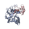


 PDBj
PDBj
 Assembly
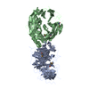


 Sample preparation
 / Beamline: 31-ID / Wavelength: 0.97929 Å
 Processing