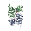
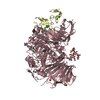
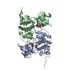
- PDB-4q69: Crystal structure of a SusD homolog (BT2259) from Bacteroides the... -
+
Open data
ID or keywords:
Loading...
-
Basic information
Entry
Database: PDB / ID: 4q69
Title
Crystal structure of a SusD homolog (BT2259) from Bacteroides thetaiotaomicron VPI-5482 at 2.50 A resolution
Components
Putative lipoprotein
Keywords
SUGAR BINDING PROTEIN / SusD-like_2 family / PF12771 / Structural Genomics / Joint Center for Structural Genomics / JCSG / Protein Structure Initiative / PSI-BIOLOGY
Mass: 18.015 Da / Num. of mol.: 292 / Source method: isolated from a natural source / Formula: H2O
Has protein modification
Y
Sequence details
THIS CONSTRUCT WAS EXPRESSED WITH AN N-TERMINAL PURIFICATION TAG MGSDKIHHHHHHENLYFQG. THE TAG WAS ...THIS CONSTRUCT WAS EXPRESSED WITH AN N-TERMINAL PURIFICATION TAG MGSDKIHHHHHHENLYFQG. THE TAG WAS REMOVED WITH TEV PROTEASE LEAVING ONLY A GLYCINE (0) FOLLOWED BY RESIDUES 28-488 OF THE TARGET SEQUENCE.
-
Experimental details
-
Experiment
Experiment
Method: X-RAY DIFFRACTION / Number of used crystals: 1
-
Sample preparation
Crystal
Density Matthews: 3.49 Å3/Da / Density % sol: 64.74 %
Crystal grow
Temperature: 277 K / Method: vapor diffusion, sitting drop / pH: 6.5 Details: 0.1M sodium citrate pH 6.5, NANODROP, VAPOR DIFFUSION, SITTING DROP, temperature 277K
Resolution: 2.5→89.672 Å / Num. all: 51369 / Num. obs: 51369 / % possible obs: 100 % / Redundancy: 6.2 % / Rsym value: 0.155 / Net I/σ(I): 9
Reflection shell
Rmerge(I) obs: 0.01 / Diffraction-ID: 1
Resolution (Å)
Redundancy (%)
Mean I/σ(I) obs
Num. measured all
Num. unique all
Rsym value
% possible all
2.5-2.56
6.3
1.9
23643
3759
1.01
100
2.56-2.64
6.3
2.1
22941
3640
0.899
100
2.64-2.71
6.3
2.4
22387
3558
0.76
100
2.71-2.8
6.3
2.9
21907
3483
0.631
100
2.8-2.89
6.3
3.8
21098
3347
0.46
100
2.89-2.99
6.3
4.3
20516
3266
0.388
100
2.99-3.1
6.3
5.4
19757
3145
0.301
100
3.1-3.23
6.3
6.8
19016
3015
0.237
100
3.23-3.37
6.3
8.2
18211
2902
0.192
100
3.37-3.54
6.3
10.3
17595
2794
0.154
100
3.54-3.73
6.3
12.4
16667
2660
0.128
100
3.73-3.95
6.3
14.5
15747
2512
0.104
100
3.95-4.23
6.3
16.1
14817
2369
0.09
100
4.23-4.56
6.2
18.6
13686
2198
0.076
100
4.56-5
6.2
18.9
12787
2052
0.069
100
5-5.59
6.2
17.1
11464
1857
0.073
100
5.59-6.46
6.2
16.4
10175
1650
0.073
100
6.46-7.91
6.1
18.1
8497
1400
0.061
100
7.91-11.18
5.9
24.4
6609
1113
0.043
100
11.18-89.672
5.4
20.9
3505
649
0.043
98.1
-
Phasing
Phasing
Method: MAD
-
Processing
Software
Name
Version
Classification
NB
MolProbity
3beta29
modelbuilding
PDB_EXTRACT
3.1
dataextraction
SHELX
phasing
SHARP
phasing
SCALA
3.3.20
datascaling
REFMAC
5.7.0032
refinement
MOSFLM
datareduction
SHELXD
phasing
Refinement
Method to determine structure: MAD / Resolution: 2.5→89.672 Å / Cor.coef. Fo:Fc: 0.957 / Cor.coef. Fo:Fc free: 0.943 / Occupancy max: 1 / Occupancy min: 0.4 / SU B: 13.373 / SU ML: 0.145 / Cross valid method: THROUGHOUT / σ(F): 0 / ESU R: 0.277 / ESU R Free: 0.199 Stereochemistry target values: MAXIMUM LIKELIHOOD WITH PHASES Details: 1. HYDROGENS HAVE BEEN ADDED IN THE RIDING POSITIONS. 2. ATOM RECORDS CONTAIN SUM OF TLS AND RESIDUAL B FACTORS. 3. ANISOU RECORDS CONTAIN SUM OF TLS AND RESIDUAL U FACTORS. 4. WATERS WERE ...Details: 1. HYDROGENS HAVE BEEN ADDED IN THE RIDING POSITIONS. 2. ATOM RECORDS CONTAIN SUM OF TLS AND RESIDUAL B FACTORS. 3. ANISOU RECORDS CONTAIN SUM OF TLS AND RESIDUAL U FACTORS. 4. WATERS WERE EXCLUDED FROM AUTOMATIC TLS ASSIGNMENT. 5. A MET-INHIBITION PROTOCOL WAS USED FOR SELENOMETHIONINE INCORPORATION DURING PROTEIN EXPRESSION. THE OCCUPANCY OF THE SE ATOMS IN THE MSE RESIDUES WAS REDUCED TO 0.75 FOR THE REDUCED SCATTERING POWER DUE TO PARTIAL S-MET INCORPORATION. 6. CHLORIDE ION (CL), SODIUM IONS (NA), AND GLYCEROL (GOL) MOLECULE FROM PURIFICATION/CRYSTALLIZATION/CRYOPROTECTION SOLUTIONS ARE MODELED.
Rfactor
Num. reflection
% reflection
Selection details
Rfree
0.1989
2605
5.1 %
RANDOM
Rwork
0.172
-
-
-
obs
0.1733
51309
99.83 %
-
Solvent computation
Ion probe radii: 0.8 Å / Shrinkage radii: 0.8 Å / VDW probe radii: 1.2 Å / Solvent model: BABINET MODEL WITH MASK
In the structure databanks used in Yorodumi, some data are registered as the other names, "COVID-19 virus" and "2019-nCoV". Here are the details of the virus and the list of structure data.
Jan 31, 2019. EMDB accession codes are about to change! (news from PDBe EMDB page)
EMDB accession codes are about to change! (news from PDBe EMDB page)
The allocation of 4 digits for EMDB accession codes will soon come to an end. Whilst these codes will remain in use, new EMDB accession codes will include an additional digit and will expand incrementally as the available range of codes is exhausted. The current 4-digit format prefixed with “EMD-” (i.e. EMD-XXXX) will advance to a 5-digit format (i.e. EMD-XXXXX), and so on. It is currently estimated that the 4-digit codes will be depleted around Spring 2019, at which point the 5-digit format will come into force.
The EM Navigator/Yorodumi systems omit the EMD- prefix.
Related info.:Q: What is EMD? / ID/Accession-code notation in Yorodumi/EM Navigator
Yorodumi is a browser for structure data from EMDB, PDB, SASBDB, etc.
This page is also the successor to EM Navigator detail page, and also detail information page/front-end page for Omokage search.
The word "yorodu" (or yorozu) is an old Japanese word meaning "ten thousand". "mi" (miru) is to see.
Related info.:EMDB / PDB / SASBDB / Comparison of 3 databanks / Yorodumi Search / Aug 31, 2016. New EM Navigator & Yorodumi / Yorodumi Papers / Jmol/JSmol / Function and homology information / Changes in new EM Navigator and Yorodumi
 Movie
Movie Controller

 Yorodumi
 Open data
 Basic information
 Components
 Keywords
 Function and homology information
 Bacteroides thetaiotaomicron (bacteria)
 X-RAY DIFFRACTION /
  Authors
 Citation
 Structure visualization
 Downloads & links
 Other downloads


 PDBj
PDBj


 Assembly

 Mass: 22.990 Da / Num. of mol.: 2 / Source method: obtained synthetically / Formula: Na
 Mass: 35.453 Da / Num. of mol.: 1 / Source method: obtained synthetically / Formula: Cl
 Mass: 92.094 Da / Num. of mol.: 1 / Source method: obtained synthetically / Formula: C3H8O3
 Mass: 18.015 Da / Num. of mol.: 292 / Source method: isolated from a natural source / Formula: H2O
 Sample preparation
 / Beamline: BL9-2 / Wavelength: 0.95369,0.97936,0.97918
 Processing