 Movie
Movie Controller

[English] 日本語
 Yorodumi
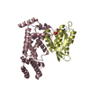
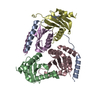
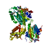
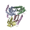
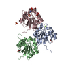
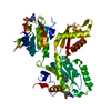
- PDB-4q5h: Shigella Effector Kinase OspG bound to AMPPNP and E2-Ub UbcH7-Ub ... -
+ Open data

- Basic information
| Entry | Database: PDB / ID: 4q5h | ||||||
|---|---|---|---|---|---|---|---|
| Title | Shigella Effector Kinase OspG bound to AMPPNP and E2-Ub UbcH7-Ub Conjugate | ||||||
 Components Components |
| ||||||
 Keywords Keywords | UNKNOWN FUNCTION / PROTEIN BINDING / protein-protein complex / Structural Genomics / Montreal-Kingston Bacterial Structural Genomics Initiative / BSGI / kinase fold / inhibition of NF-kB pathway | ||||||
| Function / homology |  Function and homology information Function and homology information: / RAS processing / Regulation of PTEN localization / : / UCH proteinases / : / Aggrephagy / Pexophagy / Transferases; Transferring phosphorus-containing groups / : ...: / RAS processing / Regulation of PTEN localization / : / UCH proteinases / : / Aggrephagy / Pexophagy / Transferases; Transferring phosphorus-containing groups / : / PINK1-PRKN Mediated Mitophagy / : / Peroxisomal protein import / cell cycle phase transition / : / Endosomal Sorting Complex Required For Transport (ESCRT) / ubiquitin-protein transferase activator activity / : / Translesion synthesis by REV1 / : / : / : / : / : / Recruitment and ATM-mediated phosphorylation of repair and signaling proteins at DNA double strand breaks / protein K11-linked ubiquitination / : / cellular response to glucocorticoid stimulus / Regulation of PTEN stability and activity / E2 ubiquitin-conjugating enzyme / cellular response to steroid hormone stimulus / CDK-mediated phosphorylation and removal of Cdc6 / FBXL7 down-regulates AURKA during mitotic entry and in early mitosis / KEAP1-NFE2L2 pathway / Neddylation / Formation of TC-NER Pre-Incision Complex / Ubiquitin-Mediated Degradation of Phosphorylated Cdc25A / Orc1 removal from chromatin / MAPK6/MAPK4 signaling / SRP-dependent cotranslational protein targeting to membrane / GTP hydrolysis and joining of the 60S ribosomal subunit / Gap-filling DNA repair synthesis and ligation in TC-NER / Formation of a pool of free 40S subunits / Nonsense Mediated Decay (NMD) independent of the Exon Junction Complex (EJC) / Nonsense Mediated Decay (NMD) enhanced by the Exon Junction Complex (EJC) / ubiquitin conjugating enzyme activity / Antigen processing: Ubiquitination & Proteasome degradation / L13a-mediated translational silencing of Ceruloplasmin expression / Dual incision in TC-NER / Ub-specific processing proteases / ubiquitin ligase complex / protein modification process / positive regulation of protein ubiquitination / Synthesis of active ubiquitin: roles of E1 and E2 enzymes / PINK1-PRKN Mediated Mitophagy / Regulation of TNFR1 signaling / Regulation of necroptotic cell death / modification-dependent protein catabolic process / protein tag activity / protein polyubiquitination / kinase activity / ubiquitin-protein transferase activity / protein autophosphorylation / peroxisome / host cell / Antigen processing: Ubiquitination & Proteasome degradation / E3 ubiquitin ligases ubiquitinate target proteins / ubiquitin-dependent protein catabolic process / transcription coactivator activity / cell population proliferation / protein ubiquitination / ubiquitin protein ligase binding / regulation of DNA-templated transcription / enzyme binding / : / RNA binding / nucleoplasm / ATP binding / nucleus / cytoplasm / cytosol Similarity search - Function | ||||||
| Biological species |  Shigella sonnei (bacteria) Shigella sonnei (bacteria)  Homo sapiens (human) Homo sapiens (human) | ||||||
| Method |  X-RAY DIFFRACTION / SYNCHROTRON / FOURIER SYNTHESIS / Resolution: 1.999 Å X-RAY DIFFRACTION / SYNCHROTRON / FOURIER SYNTHESIS / Resolution: 1.999 Å | ||||||
 Authors Authors | Cygler, M. / Grishin, A.M. / Montreal-Kingston Bacterial Structural Genomics Initiative (BSGI) | ||||||
 Citation Citation | Journal: Structure / Year: 2014 Title: Structural Basis for the Inhibition of Host Protein Ubiquitination by Shigella Effector Kinase OspG. Authors: Grishin, A.M. / Condos, T.E. / Barber, K.R. / Campbell-Valois, F.X. / Parsot, C. / Shaw, G.S. / Cygler, M. | ||||||
| History |
|
- Structure visualization
| Structure viewer | Molecule: MolmilJmol/JSmol |
|---|
- Downloads & links
-Download
| PDBx/mmCIF format | 4q5h.cif.gz | 194.1 KB | Display | PDBx/mmCIF format |
|---|---|---|---|---|
| PDB format | pdb4q5h.ent.gz | 152.8 KB | Display | PDB format |
| PDBx/mmJSON format | 4q5h.json.gz | Tree view | PDBx/mmJSON format | |
| Others |  Other downloads Other downloads |
-Validation report
| Arichive directory | https://data.pdbj.org/pub/pdb/validation_reports/q5/4q5hftp://data.pdbj.org/pub/pdb/validation_reports/q5/4q5h | HTTPS FTP |
|---|
-Related structure data
| Related structure data |  4q5eC  4qseS S: Starting model for refinement C: citing same article ( |
|---|---|
| Similar structure data |


-Links
 PDBj
PDBj


- Assembly
| Deposited unit | 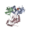
| ||||||||
|---|---|---|---|---|---|---|---|---|---|
| 1 |
| ||||||||
| Unit cell |
|
-Components
-Protein , 3 types, 3 molecules ABC
| #1: Protein | Mass: 20116.547 Da / Num. of mol.: 1 / Fragment: unp residues 26-193 Source method: isolated from a genetically manipulated source Source: (gene. exp.) Shigella sonnei (bacteria) / Strain: 2457T / Gene: ospG, SSON_P170 / Plasmid: pMCSG7 / Production host: References: UniProt: Q3YTH2, Transferases; Transferring phosphorus-containing groups |
|---|---|
| #2: Protein | Mass: 8642.873 Da / Num. of mol.: 1 / Mutation: C17S, C137S Source method: isolated from a genetically manipulated source Source: (gene. exp.) Strain: ATCC 204508 / S288c / Gene: SCD2, UBE2L3, UBI4, YLL039C / Plasmid: pET28a / Production host: |
| #3: Protein | Mass: 18052.662 Da / Num. of mol.: 1 / Mutation: K48R, G76C Source method: isolated from a genetically manipulated source Source: (gene. exp.) Homo sapiens (human) / Gene: UBCE7, UBCH7, UBE2L3, UBI4 / Plasmid: pET3a / Production host: |
-Non-polymers , 3 types, 232 molecules 


| #4: Chemical | ChemComp-MG /  Mass: 24.305 Da / Num. of mol.: 1 / Source method: obtained synthetically / Formula: Mg Mass: 24.305 Da / Num. of mol.: 1 / Source method: obtained synthetically / Formula: Mg |
|---|---|
| #5: Chemical | ChemComp-ANP /  Mass: 506.196 Da / Num. of mol.: 1 / Source method: obtained synthetically / Formula: C10H17N6O12P3 / Comment: AMP-PNP, energy-carrying molecule analogue*YM Mass: 506.196 Da / Num. of mol.: 1 / Source method: obtained synthetically / Formula: C10H17N6O12P3 / Comment: AMP-PNP, energy-carrying molecule analogue*YM |
| #6: Water | ChemComp-HOH / Mass: 18.015 Da / Num. of mol.: 230 / Source method: isolated from a natural source / Formula: H2O |
-Details
| Has protein modification | Y |
|---|
-Experimental details
-Experiment
| Experiment | Method: X-RAY DIFFRACTION / Number of used crystals: 1 |
|---|
- Sample preparation
| Crystal | Density Matthews: 2.64 Å3/Da / Density % sol: 53.4 % |
|---|---|
| Crystal grow | Temperature: 293 K / Method: vapor diffusion, hanging drop / pH: 8 Details: 100mM Hepes 8.2, 15% PEG 4000, 100 mM MgCl2, pH 8.0, VAPOR DIFFUSION, HANGING DROP, temperature 293K |
-Data collection
| Diffraction | Mean temperature: 77.2 K | |||||||||||||||||||||||||||||||||||||||||||||||||||||||||||||||||||||||||||||||||||||||||||||||||||||||||
|---|---|---|---|---|---|---|---|---|---|---|---|---|---|---|---|---|---|---|---|---|---|---|---|---|---|---|---|---|---|---|---|---|---|---|---|---|---|---|---|---|---|---|---|---|---|---|---|---|---|---|---|---|---|---|---|---|---|---|---|---|---|---|---|---|---|---|---|---|---|---|---|---|---|---|---|---|---|---|---|---|---|---|---|---|---|---|---|---|---|---|---|---|---|---|---|---|---|---|---|---|---|---|---|---|---|---|
| Diffraction source | Source: SYNCHROTRON / Site: CLSI  / Beamline: 08ID-1 / Wavelength: 0.97949 Å / Beamline: 08ID-1 / Wavelength: 0.97949 Å | |||||||||||||||||||||||||||||||||||||||||||||||||||||||||||||||||||||||||||||||||||||||||||||||||||||||||
| Detector | Type: RAYONIX MX-300 / Detector: CCD / Date: Feb 13, 2013 | |||||||||||||||||||||||||||||||||||||||||||||||||||||||||||||||||||||||||||||||||||||||||||||||||||||||||
| Radiation | Monochromator: dcm WITH CRYO-COOLED 1ST CRYSTAL SAGITALLY BENT 2ND CRYSTAL FOLLOWED BY VERTICALLY FOCUSING MIRROR Protocol: SINGLE WAVELENGTH / Monochromatic (M) / Laue (L): M / Scattering type: x-ray | |||||||||||||||||||||||||||||||||||||||||||||||||||||||||||||||||||||||||||||||||||||||||||||||||||||||||
| Radiation wavelength | Wavelength: 0.97949 Å / Relative weight: 1 | |||||||||||||||||||||||||||||||||||||||||||||||||||||||||||||||||||||||||||||||||||||||||||||||||||||||||
| Reflection | Resolution: 2→50 Å / Num. obs: 33962 / % possible obs: 99.9 % / Redundancy: 7.1 % / Biso Wilson estimate: 35.08 Å2 / Rmerge(I) obs: 0.094 / Net I/σ(I): 11 | |||||||||||||||||||||||||||||||||||||||||||||||||||||||||||||||||||||||||||||||||||||||||||||||||||||||||
| Reflection shell |
|
- Processing
| Software |
| ||||||||||||||||||||||||||||||||||||||||||||||||||||||||||||||||||||||||||||||||||||||||||||||||||||||||||||||||||||||||||||||||||||||||||||||||||||||
|---|---|---|---|---|---|---|---|---|---|---|---|---|---|---|---|---|---|---|---|---|---|---|---|---|---|---|---|---|---|---|---|---|---|---|---|---|---|---|---|---|---|---|---|---|---|---|---|---|---|---|---|---|---|---|---|---|---|---|---|---|---|---|---|---|---|---|---|---|---|---|---|---|---|---|---|---|---|---|---|---|---|---|---|---|---|---|---|---|---|---|---|---|---|---|---|---|---|---|---|---|---|---|---|---|---|---|---|---|---|---|---|---|---|---|---|---|---|---|---|---|---|---|---|---|---|---|---|---|---|---|---|---|---|---|---|---|---|---|---|---|---|---|---|---|---|---|---|---|---|---|---|
| Refinement | Method to determine structure: FOURIER SYNTHESIS Starting model: pdb entry 4QSE Resolution: 1.999→44.939 Å / SU ML: 0.24 / σ(F): 1.33 / Phase error: 24.73 / Stereochemistry target values: ML
| ||||||||||||||||||||||||||||||||||||||||||||||||||||||||||||||||||||||||||||||||||||||||||||||||||||||||||||||||||||||||||||||||||||||||||||||||||||||
| Solvent computation | Shrinkage radii: 0.9 Å / VDW probe radii: 1.11 Å / Solvent model: FLAT BULK SOLVENT MODEL | ||||||||||||||||||||||||||||||||||||||||||||||||||||||||||||||||||||||||||||||||||||||||||||||||||||||||||||||||||||||||||||||||||||||||||||||||||||||
| Displacement parameters | Biso mean: 53.66 Å2 | ||||||||||||||||||||||||||||||||||||||||||||||||||||||||||||||||||||||||||||||||||||||||||||||||||||||||||||||||||||||||||||||||||||||||||||||||||||||
| Refinement step | Cycle: LAST / Resolution: 1.999→44.939 Å
| ||||||||||||||||||||||||||||||||||||||||||||||||||||||||||||||||||||||||||||||||||||||||||||||||||||||||||||||||||||||||||||||||||||||||||||||||||||||
| Refine LS restraints |
| ||||||||||||||||||||||||||||||||||||||||||||||||||||||||||||||||||||||||||||||||||||||||||||||||||||||||||||||||||||||||||||||||||||||||||||||||||||||
| LS refinement shell |
| ||||||||||||||||||||||||||||||||||||||||||||||||||||||||||||||||||||||||||||||||||||||||||||||||||||||||||||||||||||||||||||||||||||||||||||||||||||||
| Refinement TLS params. | Method: refined / Refine-ID: X-RAY DIFFRACTION
| ||||||||||||||||||||||||||||||||||||||||||||||||||||||||||||||||||||||||||||||||||||||||||||||||||||||||||||||||||||||||||||||||||||||||||||||||||||||
| Refinement TLS group |
|
