 Movie
Movie Controller

[English] 日本語
 Yorodumi
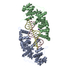
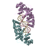
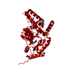
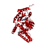
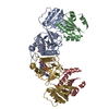
- PDB-4q0w: he catalytic core of Rad2 in complex with DNA substrate (complex II) -
+ Open data

- Basic information
| Entry | Database: PDB / ID: 4q0w | ||||||
|---|---|---|---|---|---|---|---|
| Title | he catalytic core of Rad2 in complex with DNA substrate (complex II) | ||||||
 Components Components |
| ||||||
 Keywords Keywords | Hydrolase/DNA / ba Rossmann-like / DNA repair / TFIIH / nucleus / Hydrolase-DNA complex | ||||||
| Function / homology |  Function and homology information Function and homology informationsingle-stranded DNA endonuclease activity / nucleotide-excision repair factor 3 complex / Dual incision in TC-NER / nucleotide-excision repair / DNA endonuclease activity / single-stranded DNA binding / transcription by RNA polymerase II / Hydrolases; Acting on ester bonds / metal ion binding / nucleus Similarity search - Function | ||||||
| Biological species |  | ||||||
| Method |  X-RAY DIFFRACTION / SYNCHROTRON / MOLECULAR REPLACEMENT / Resolution: 2.1 Å X-RAY DIFFRACTION / SYNCHROTRON / MOLECULAR REPLACEMENT / Resolution: 2.1 Å | ||||||
 Authors Authors | Mietus, M. / Nowak, E. / Jaciuk, M. / Kustosz, P. / Nowotny, M. | ||||||
 Citation Citation | Journal: Nucleic Acids Res. / Year: 2014 Title: Crystal structure of the catalytic core of Rad2: insights into the mechanism of substrate binding. Authors: Mietus, M. / Nowak, E. / Jaciuk, M. / Kustosz, P. / Studnicka, J. / Nowotny, M. | ||||||
| History |
|
- Structure visualization
| Structure viewer | Molecule: MolmilJmol/JSmol |
|---|
- Downloads & links
-Download
| PDBx/mmCIF format | 4q0w.cif.gz | 172.2 KB | Display | PDBx/mmCIF format |
|---|---|---|---|---|
| PDB format | pdb4q0w.ent.gz | 130 KB | Display | PDB format |
| PDBx/mmJSON format | 4q0w.json.gz | Tree view | PDBx/mmJSON format | |
| Others |  Other downloads Other downloads |
-Validation report
| Arichive directory | https://data.pdbj.org/pub/pdb/validation_reports/q0/4q0wftp://data.pdbj.org/pub/pdb/validation_reports/q0/4q0w | HTTPS FTP |
|---|
-Related structure data
| Related structure data |  4q0rSC  4q0zC  4q10C S: Starting model for refinement C: citing same article ( |
|---|---|
| Similar structure data |


-Links
 PDBj
PDBj


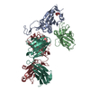
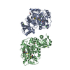
- Assembly
| Deposited unit | 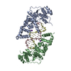
| ||||||||
|---|---|---|---|---|---|---|---|---|---|
| 1 |
| ||||||||
| Unit cell |
|
-Components
-Protein , 1 types, 2 molecules AB
| #1: Protein | Mass: 42189.062 Da / Num. of mol.: 2 / Fragment: Rad2 Source method: isolated from a genetically manipulated source Source: (gene. exp.) Strain: ATCC 204508 / S288c / Gene: RAD2, YGR258C / Production host:  References: UniProt: P07276, Hydrolases; Acting on ester bonds |
|---|
-DNA chain , 2 types, 2 molecules CD
| #2: DNA chain | Mass: 5408.500 Da / Num. of mol.: 1 / Source method: obtained synthetically |
|---|---|
| #3: DNA chain | Mass: 5586.625 Da / Num. of mol.: 1 / Source method: obtained synthetically |
-Non-polymers , 3 types, 571 molecules 


| #4: Chemical |  Mass: 40.078 Da / Num. of mol.: 2 / Source method: obtained synthetically / Formula: Ca Mass: 40.078 Da / Num. of mol.: 2 / Source method: obtained synthetically / Formula: Ca#5: Chemical |  Mass: 39.098 Da / Num. of mol.: 2 / Source method: obtained synthetically / Formula: K Mass: 39.098 Da / Num. of mol.: 2 / Source method: obtained synthetically / Formula: K#6: Water | ChemComp-HOH / | Mass: 18.015 Da / Num. of mol.: 567 / Source method: isolated from a natural source / Formula: H2O |
|---|
-Experimental details
-Experiment
| Experiment | Method: X-RAY DIFFRACTION / Number of used crystals: 1 |
|---|
- Sample preparation
| Crystal | Density Matthews: 2.76 Å3/Da / Density % sol: 55.41 % |
|---|---|
| Crystal grow | Temperature: 291 K / Method: vapor diffusion, sitting drop / pH: 7.5 Details: 9% (w/v) PEG 20000, 20% (v/v) PEG MME 550, 0.2 M D-glucose, 0.2 M D-mannose, 0.2 M D-galactose, 0.2 M L-fructose, 0.2 M D-xylose, 0.2 M N-acetyl-D-glucosamine, and 0.1 M MOPS/HEPES-Na, pH 7. ...Details: 9% (w/v) PEG 20000, 20% (v/v) PEG MME 550, 0.2 M D-glucose, 0.2 M D-mannose, 0.2 M D-galactose, 0.2 M L-fructose, 0.2 M D-xylose, 0.2 M N-acetyl-D-glucosamine, and 0.1 M MOPS/HEPES-Na, pH 7.5, VAPOR DIFFUSION, SITTING DROP, temperature 291K |
-Data collection
| Diffraction | Mean temperature: 100 K |
|---|---|
| Diffraction source | Source: SYNCHROTRON / Site: BESSY  / Beamline: 14.2 / Wavelength: 0.8944 Å / Beamline: 14.2 / Wavelength: 0.8944 Å |
| Detector | Type: RAYONIX MX-225 / Detector: CCD / Date: May 23, 2013 |
| Radiation | Monochromator: double crystal / Protocol: SINGLE WAVELENGTH / Monochromatic (M) / Laue (L): M / Scattering type: x-ray |
| Radiation wavelength | Wavelength: 0.8944 Å / Relative weight: 1 |
| Reflection | Resolution: 2.14→40 Å / Num. all: 62290 / Num. obs: 62159 / % possible obs: 99.79 % / Observed criterion σ(F): 2.1 / Observed criterion σ(I): 2.1 / Redundancy: 7.1 % / Rmerge(I) obs: 0.088 |
| Reflection shell | Resolution: 2.1→2.14 Å / Redundancy: 6.3 % / Rmerge(I) obs: 0.831 / Mean I/σ(I) obs: 2.1 / % possible all: 99.3 |
- Processing
| Software |
| ||||||||||||||||||||||||||||||||||||||||||||||||||||||||||||||||||||||||||||||||||||||||||||||||||||||||||||||||||||||||||||||||||||||||||||||||||||||||||||||||||||||||
|---|---|---|---|---|---|---|---|---|---|---|---|---|---|---|---|---|---|---|---|---|---|---|---|---|---|---|---|---|---|---|---|---|---|---|---|---|---|---|---|---|---|---|---|---|---|---|---|---|---|---|---|---|---|---|---|---|---|---|---|---|---|---|---|---|---|---|---|---|---|---|---|---|---|---|---|---|---|---|---|---|---|---|---|---|---|---|---|---|---|---|---|---|---|---|---|---|---|---|---|---|---|---|---|---|---|---|---|---|---|---|---|---|---|---|---|---|---|---|---|---|---|---|---|---|---|---|---|---|---|---|---|---|---|---|---|---|---|---|---|---|---|---|---|---|---|---|---|---|---|---|---|---|---|---|---|---|---|---|---|---|---|---|---|---|---|---|---|---|---|
| Refinement | Method to determine structure: MOLECULAR REPLACEMENT Starting model: pdb entry 4Q0R Resolution: 2.1→30.386 Å / SU ML: 0.23 / σ(F): 1.34 / Phase error: 23.31 / Stereochemistry target values: ML
| ||||||||||||||||||||||||||||||||||||||||||||||||||||||||||||||||||||||||||||||||||||||||||||||||||||||||||||||||||||||||||||||||||||||||||||||||||||||||||||||||||||||||
| Solvent computation | Shrinkage radii: 0.9 Å / VDW probe radii: 1.11 Å / Solvent model: FLAT BULK SOLVENT MODEL | ||||||||||||||||||||||||||||||||||||||||||||||||||||||||||||||||||||||||||||||||||||||||||||||||||||||||||||||||||||||||||||||||||||||||||||||||||||||||||||||||||||||||
| Refinement step | Cycle: LAST / Resolution: 2.1→30.386 Å
| ||||||||||||||||||||||||||||||||||||||||||||||||||||||||||||||||||||||||||||||||||||||||||||||||||||||||||||||||||||||||||||||||||||||||||||||||||||||||||||||||||||||||
| Refine LS restraints |
| ||||||||||||||||||||||||||||||||||||||||||||||||||||||||||||||||||||||||||||||||||||||||||||||||||||||||||||||||||||||||||||||||||||||||||||||||||||||||||||||||||||||||
| LS refinement shell |
|
