| Deposited unit | A: 3-Hydroxyacyl-CoA dehydrogenase
B: 3-Hydroxyacyl-CoA dehydrogenase
C: 3-Hydroxyacyl-CoA dehydrogenase
D: 3-Hydroxyacyl-CoA dehydrogenase
E: 3-Hydroxyacyl-CoA dehydrogenase
F: 3-Hydroxyacyl-CoA dehydrogenase
G: 3-Hydroxyacyl-CoA dehydrogenase
H: 3-Hydroxyacyl-CoA dehydrogenase
I: 3-Hydroxyacyl-CoA dehydrogenase
hetero molecules
| Theoretical mass | Number of molelcules |
|---|
| Total (without water) | 278,305 | 18 |
|---|
| Polymers | 272,334 | 9 |
|---|
| Non-polymers | 5,971 | 9 |
|---|
| Water | 1,820 | 101 |
|---|
|
|---|
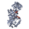
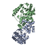
| 1 | A: 3-Hydroxyacyl-CoA dehydrogenase
hetero molecules
A: 3-Hydroxyacyl-CoA dehydrogenase
hetero molecules
| Theoretical mass | Number of molelcules |
|---|
| Total (without water) | 61,845 | 4 |
|---|
| Polymers | 60,519 | 2 |
|---|
| Non-polymers | 1,327 | 2 |
|---|
| Water | 36 | 2 |
|---|
| Type | Name | Symmetry operation | Number |
|---|
| identity operation | 1_555 | x,y,z | 1 | | crystal symmetry operation | 2_554 | -x,y,-z-1 | 1 |
| Buried area | 3720 Å2 |
|---|
| ΔGint | -27 kcal/mol |
|---|
| Surface area | 24580 Å2 |
|---|
| Method | PISA |
|---|
|
|---|
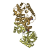
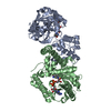
| 2 | B: 3-Hydroxyacyl-CoA dehydrogenase
C: 3-Hydroxyacyl-CoA dehydrogenase
hetero molecules
| Theoretical mass | Number of molelcules |
|---|
| Total (without water) | 61,845 | 4 |
|---|
| Polymers | 60,519 | 2 |
|---|
| Non-polymers | 1,327 | 2 |
|---|
| Water | 36 | 2 |
|---|
| Type | Name | Symmetry operation | Number |
|---|
| identity operation | 1_555 | x,y,z | 1 |
| Buried area | 3770 Å2 |
|---|
| ΔGint | -26 kcal/mol |
|---|
| Surface area | 23580 Å2 |
|---|
| Method | PISA |
|---|
|
|---|
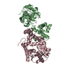
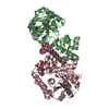
| 3 | D: 3-Hydroxyacyl-CoA dehydrogenase
G: 3-Hydroxyacyl-CoA dehydrogenase
hetero molecules
| Theoretical mass | Number of molelcules |
|---|
| Total (without water) | 61,845 | 4 |
|---|
| Polymers | 60,519 | 2 |
|---|
| Non-polymers | 1,327 | 2 |
|---|
| Water | 36 | 2 |
|---|
| Type | Name | Symmetry operation | Number |
|---|
| identity operation | 1_555 | x,y,z | 1 |
| Buried area | 3770 Å2 |
|---|
| ΔGint | -25 kcal/mol |
|---|
| Surface area | 24410 Å2 |
|---|
| Method | PISA |
|---|
|
|---|
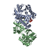
| 4 | E: 3-Hydroxyacyl-CoA dehydrogenase
F: 3-Hydroxyacyl-CoA dehydrogenase
hetero molecules
| Theoretical mass | Number of molelcules |
|---|
| Total (without water) | 61,845 | 4 |
|---|
| Polymers | 60,519 | 2 |
|---|
| Non-polymers | 1,327 | 2 |
|---|
| Water | 36 | 2 |
|---|
| Type | Name | Symmetry operation | Number |
|---|
| identity operation | 1_555 | x,y,z | 1 |
| Buried area | 3750 Å2 |
|---|
| ΔGint | -27 kcal/mol |
|---|
| Surface area | 23490 Å2 |
|---|
| Method | PISA |
|---|
|
|---|
| 5 | H: 3-Hydroxyacyl-CoA dehydrogenase
I: 3-Hydroxyacyl-CoA dehydrogenase
hetero molecules
| Theoretical mass | Number of molelcules |
|---|
| Total (without water) | 61,845 | 4 |
|---|
| Polymers | 60,519 | 2 |
|---|
| Non-polymers | 1,327 | 2 |
|---|
| Water | 36 | 2 |
|---|
| Type | Name | Symmetry operation | Number |
|---|
| identity operation | 1_555 | x,y,z | 1 |
| Buried area | 3760 Å2 |
|---|
| ΔGint | -27 kcal/mol |
|---|
| Surface area | 23480 Å2 |
|---|
| Method | PISA |
|---|
|
|---|
| Unit cell | | Length a, b, c (Å) | 235.076, 135.586, 97.445 |
|---|
| Angle α, β, γ (deg.) | 90.00, 90.09, 90.00 |
|---|
| Int Tables number | 5 |
|---|
| Space group name H-M | C121 |
|---|
|
|---|
 Movie
Movie Controller

 Yorodumi
 Open data
 Basic information
 Components
 Keywords
 Function and homology information
 Ralstonia eutropha H16 (bacteria)
 X-RAY DIFFRACTION /
  Authors
 Citation
 Structure visualization
 Downloads & links
 Other downloads


 PDBj
PDBj


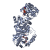
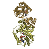
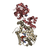
 Assembly
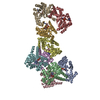


 Mass: 663.425 Da / Num. of mol.: 9 / Source method: obtained synthetically / Formula: C21H27N7O14P2 / Comment: NAD*YM
 Mass: 18.015 Da / Num. of mol.: 101 / Source method: isolated from a natural source / Formula: H2O
 Sample preparation
 / Beamline: 7A (6B, 6C1) / Wavelength: 1 Å
 Processing