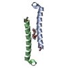
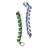
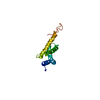
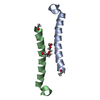
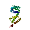
[English] 日本語
Yorodumi
- PDB-4pz5: Crystal structure of the second and third fibronectin F1 modules ... -

+
Open data


ID or keywords:

Loading...

-
Basic information

Entry
Database: PDB / ID: 4pz5
TitleCrystal structure of the second and third fibronectin F1 modules in complex with a fragment of BBK32 from Borrelia burgdorferi
Components
  • Fibronectin
  • Fibronectin-binding protein BBK32
KeywordsCELL ADHESION / Fibronectin type one / Bacterial adhesion / Fibronectin binding / Extracellular matrix / Plasma
Function / homology
Function and homology information


negative regulation of monocyte activation / calcium-independent cell-matrix adhesion / negative regulation of transforming growth factor beta production / Fibronectin matrix formation / Extracellular matrix organization / positive regulation of substrate-dependent cell migration, cell attachment to substrate / neural crest cell migration involved in autonomic nervous system development / peptidase activator activity / fibrinogen complex / peptide cross-linking ...negative regulation of monocyte activation / calcium-independent cell-matrix adhesion / negative regulation of transforming growth factor beta production / Fibronectin matrix formation / Extracellular matrix organization / positive regulation of substrate-dependent cell migration, cell attachment to substrate / neural crest cell migration involved in autonomic nervous system development / peptidase activator activity / fibrinogen complex / peptide cross-linking / integrin activation / ALK mutants bind TKIs / cell-substrate junction assembly / biological process involved in interaction with symbiont / proteoglycan binding / Molecules associated with elastic fibres / MET activates PTK2 signaling / extracellular matrix structural constituent / Syndecan interactions / p130Cas linkage to MAPK signaling for integrins / endodermal cell differentiation / GRB2:SOS provides linkage to MAPK signaling for Integrins / fibronectin binding / endoplasmic reticulum-Golgi intermediate compartment / Non-integrin membrane-ECM interactions / basement membrane / ECM proteoglycans / Integrin cell surface interactions / positive regulation of axon extension / collagen binding / Degradation of the extracellular matrix / Integrin signaling / substrate adhesion-dependent cell spreading / regulation of ERK1 and ERK2 cascade / cell-matrix adhesion / extracellular matrix / platelet alpha granule lumen / acute-phase response / integrin-mediated signaling pathway / Cell surface interactions at the vascular wall / Post-translational protein phosphorylation / regulation of protein phosphorylation / Signaling by high-kinase activity BRAF mutants / wound healing / MAP2K and MAPK activation / response to wounding / Signaling by RAF1 mutants / Signaling by moderate kinase activity BRAF mutants / Paradoxical activation of RAF signaling by kinase inactive BRAF / GPER1 signaling / Signaling downstream of RAS mutants / Regulation of Insulin-like Growth Factor (IGF) transport and uptake by Insulin-like Growth Factor Binding Proteins (IGFBPs) / Signaling by BRAF and RAF1 fusions / positive regulation of fibroblast proliferation / Signaling by ALK fusions and activated point mutants / integrin binding / Platelet degranulation / nervous system development / heparin binding / heart development / regulation of cell shape / angiogenesis / Interleukin-4 and Interleukin-13 signaling / collagen-containing extracellular matrix / protease binding / molecular adaptor activity / positive regulation of phosphatidylinositol 3-kinase/protein kinase B signal transduction / blood microparticle / cell adhesion / apical plasma membrane / endoplasmic reticulum lumen / signaling receptor binding / positive regulation of cell population proliferation / positive regulation of gene expression / extracellular space / extracellular exosome / extracellular region / identical protein binding / plasma membrane
Similarity search - Function
Fibronectin type I domain / Fibronectin, type I / Fibronectin type-I domain signature. / Fibronectin type-I domain profile. / Fibronectin type 1 domain / Complement Module, domain 1 / Complement Module; domain 1 / Fibronectin type II domain / Fibronectin type II domain superfamily / Fibronectin type II domain ...Fibronectin type I domain / Fibronectin, type I / Fibronectin type-I domain signature. / Fibronectin type-I domain profile. / Fibronectin type 1 domain / Complement Module, domain 1 / Complement Module; domain 1 / Fibronectin type II domain / Fibronectin type II domain superfamily / Fibronectin type II domain / Fibronectin type-II collagen-binding domain signature. / Fibronectin type-II collagen-binding domain profile. / Fibronectin type 2 domain / Kringle-like fold / Fibronectin type III domain / EGF-like domain signature 1. / Fibronectin type 3 domain / Fibronectin type-III domain profile. / Fibronectin type III / Fibronectin type III superfamily / Ribbon / Prokaryotic membrane lipoprotein lipid attachment site profile. / Immunoglobulin-like fold / Mainly Beta
Similarity search - Domain/homology
Fibronectin-binding protein BBK32 / Fibronectin
Similarity search - Component
Biological speciesHomo sapiens (human)
Borrelia burgdorferi (Lyme disease spirochete)
MethodX-RAY DIFFRACTION / SYNCHROTRON / MOLECULAR REPLACEMENT / Resolution: 1.96 Å
AuthorsHarris, G. / Potts, J.R.
CitationJournal: J.Biol.Chem. / Year: 2014
Title: Borrelia burgdorferi Protein BBK32 Binds to Soluble Fibronectin via the N-terminal 70-kDa Region, Causing Fibronectin to Undergo Conformational Extension.
Authors: Harris, G. / Ma, W. / Maurer, L.M. / Potts, J.R. / Mosher, D.F.
History
DepositionMar 28, 2014Deposition site: RCSB / Processing site: RCSB
Revision 1.0Jul 2, 2014Provider: repository / Type: Initial release
Revision 1.1Jul 23, 2014Group: Database references
Revision 1.2Aug 27, 2014Group: Database references
Revision 1.3Jul 17, 2019Group: Data collection / Derived calculations / Refinement description
Category: software / struct_conn
Item: _software.classification / _software.name ..._software.classification / _software.name / _software.version / _struct_conn.pdbx_leaving_atom_flag
Revision 1.4Jan 29, 2020Group: Data collection / Database references / Category: reflns / reflns_shell / struct_ref_seq_dif
Item: _reflns.pdbx_Rmerge_I_obs / _reflns_shell.Rmerge_I_obs / _struct_ref_seq_dif.details
Revision 1.5Sep 20, 2023Group: Data collection / Database references / Refinement description
Category: chem_comp_atom / chem_comp_bond ...chem_comp_atom / chem_comp_bond / database_2 / pdbx_initial_refinement_model
Item: _database_2.pdbx_DOI / _database_2.pdbx_database_accession

-
Structure visualization

Structure viewerMolecule:
MolmilJmol/JSmol

Downloads & links

-
Assembly

Deposited unit
A: Fibronectin
B: Fibronectin-binding protein BBK32


Theoretical massNumber of molelcules
Total (without water)11,9822
Polymers11,9822
Non-polymers00
Water2,324129
1


  • Idetical with deposited unit
  • defined by author&software
TypeNameSymmetry operationNumber
identity operation1_555x,y,z1
Buried area1160 Å2
ΔGint-3 kcal/mol
Surface area6880 Å2
MethodPISA
Unit cell
Length a, b, c (Å)86.580, 86.580, 63.570
Angle α, β, γ (deg.)90.00, 90.00, 120.00
Int Tables number179
Space group name H-MP6522
Components on special symmetry positions
IDModelComponents
11A-282-

HOH

-
Components

#1: Protein Fibronectin / FN / Cold-insoluble globulin / CIG / Anastellin / Ugl-Y1 / Ugl-Y2 / Ugl-Y3


Mass: 10120.389 Da / Num. of mol.: 1 / Fragment: 2FN1-3FN1, UNP RESIDUES 93-182
Source method: isolated from a genetically manipulated source
Source: (gene. exp.) Homo sapiens (human) / Gene: FN1, FN / Production host: 
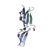
Pichia pastoris (fungus) / References: UniProt: P02751
#2: Protein/peptide Fibronectin-binding protein BBK32


Mass: 1861.823 Da / Num. of mol.: 1 / Fragment: BBK32TwL, UNP RESIDUES 175-189 / Source method: obtained synthetically
Source: (synth.) Borrelia burgdorferi (Lyme disease spirochete)
References: UniProt: O50835
#3: Water ChemComp-HOH / water


Mass: 18.015 Da / Num. of mol.: 129 / Source method: isolated from a natural source / Formula: H2O

-
Experimental details

-
Experiment

ExperimentMethod: X-RAY DIFFRACTION / Number of used crystals: 1

-
Sample preparation

CrystalDensity Matthews: 2.87 Å3/Da / Density % sol: 57.14 %
Crystal growTemperature: 291 K / Method: vapor diffusion, sitting drop / pH: 6.9
Details: 1.4 M Na/K phosphate, pH 6.9, VAPOR DIFFUSION, SITTING DROP, temperature 291K

-
Data collection

DiffractionMean temperature: 100 K
Diffraction sourceSource: SYNCHROTRON / Site: ESRF / Beamline: ID23-1 / Wavelength: 1.0039 Å
DetectorType: ADSC QUANTUM 315r / Detector: CCD / Date: Feb 23, 2007
RadiationMonochromator: Si 111 Channel / Protocol: SINGLE WAVELENGTH / Monochromatic (M) / Laue (L): M / Scattering type: x-ray
Radiation wavelengthWavelength: 1.0039 Å / Relative weight: 1
ReflectionResolution: 1.96→48.49 Å / Num. all: 10551 / Num. obs: 10524 / % possible obs: 99.8 % / Observed criterion σ(F): 0 / Observed criterion σ(I): 2 / Redundancy: 7.2 % / Biso Wilson estimate: 16.6 Å2 / Rmerge(I) obs: 0.104 / Net I/σ(I): 12.9
Reflection shellResolution: 1.96→2.03 Å / Redundancy: 7 % / Rmerge(I) obs: 0.59 / Mean I/σ(I) obs: 4.3 / Num. unique all: 1033 / % possible all: 100

-
Processing

Software
NameVersionClassification
PHENIX(phenix.refine: 1.8.4_1496)refinement
REFMACrefinement
ADSCQuantumdata collection
MOSFLMdata reduction
SCALAdata scaling
REFMACphasing
RefinementMethod to determine structure: MOLECULAR REPLACEMENT
Starting model: PDB ENTRY 2CG7
Resolution: 1.96→48.49 Å / SU ML: 0.11 / Isotropic thermal model: Isotropic / σ(F): 1.34 / σ(I): 2 / Phase error: 18.61 / Stereochemistry target values: ML
RfactorNum. reflection% reflectionSelection details
Rfree0.199 512 4.87 %Random
Rwork0.1612 ---
obs0.163 10524 99.8 %-
all-10551 --
Solvent computationShrinkage radii: 0.9 Å / VDW probe radii: 1.11 Å / Solvent model: FLAT BULK SOLVENT MODEL
Displacement parametersBiso mean: 25.9 Å2
Refinement stepCycle: LAST / Resolution: 1.96→48.49 Å
ProteinNucleic acidLigandSolventTotal
Num. atoms820 0 0 129 949
Refine LS restraints
Refine-IDTypeDev idealNumber
X-RAY DIFFRACTIONf_bond_d0.007852
X-RAY DIFFRACTIONf_angle_d0.9691147
X-RAY DIFFRACTIONf_dihedral_angle_d11.621317
X-RAY DIFFRACTIONf_chiral_restr0.041116
X-RAY DIFFRACTIONf_plane_restr0.003150
LS refinement shell
Resolution (Å)Rfactor RfreeNum. reflection RfreeRfactor RworkNum. reflection RworkRefine-ID% reflection obs (%)
1.9613-2.15860.20211040.18822454X-RAY DIFFRACTION100
2.1586-2.4710.19911450.17712440X-RAY DIFFRACTION100
2.471-3.11310.24951260.17552489X-RAY DIFFRACTION100
3.1131-48.50340.17441370.14192610X-RAY DIFFRACTION99
Refinement TLS params.

Method: refined / Refine-ID: X-RAY DIFFRACTION

IDL112)L122)L132)L222)L232)L332)S11 (Å °)S12 (Å °)S13 (Å °)S21 (Å °)S22 (Å °)S23 (Å °)S31 (Å °)S32 (Å °)S33 (Å °)T112)T122)T132)T222)T232)T332)Origin x (Å)Origin y (Å)Origin z (Å)
18.2479-2.893-2.40574.31664.0213.82480.17530.48950.1223-0.16430.0334-0.6028-0.21210.2957-0.2160.3-0.01610.04890.16640.02610.1989-28.695210.634648.4557
25.88752.64330.84065.6321.53222.00330.00220.01-0.2705-0.10630.2169-1.63170.00450.7476-0.18660.20020.00750.02820.2567-0.01070.3385-24.90515.598950.6579
33.2171-3.4317-3.36955.47564.24444.64970.1677-0.03540.2887-0.059-0.001-0.3316-0.06210.0319-0.18810.1818-0.0410.00290.12470.01470.1709-33.243410.250258.2352
41.9346-1.2253-1.75161.3958-0.4595.8751-0.04120.07210.01470.1083-0.13760.0799-0.05590.00570.12540.1491-0.0448-0.00430.10350.01560.1326-36.61298.446260.2537
52.6851-0.110.00485.77494.07533.12470.0736-0.07520.1569-0.05960.0067-0.3405-0.42210.1823-0.06940.1669-0.01610.04750.12650.03470.1822-33.447310.76677.3595
64.6809-0.5778-4.53331.43051.44694.9772-0.1451-0.27991.4569-0.4784-0.17690.2348-1.0413-0.77380.26970.3660.0652-0.01460.2012-0.09710.4022-42.496719.241488.604
70.1759-0.5872-0.63466.57085.48654.6858-0.0148-0.0125-0.03980.31480.0497-0.10230.50980.0952-0.03590.1279-0.00850.02790.08440.01160.1276-39.46277.500779.5083
82.28431.4989-2.64121.0337-1.82523.1939-0.1388-0.07280.0128-0.1603-0.0404-0.11220.33960.33560.16740.22130.01940.00030.2837-0.00380.177-42.17659.990950.9239
Refinement TLS group
IDRefine-IDRefine TLS-IDSelection details
1X-RAY DIFFRACTION1chain 'A' and (resid 63 through 72 )
2X-RAY DIFFRACTION2chain 'A' and (resid 73 through 79 )
3X-RAY DIFFRACTION3chain 'A' and (resid 80 through 96 )
4X-RAY DIFFRACTION4chain 'A' and (resid 97 through 115 )
5X-RAY DIFFRACTION5chain 'A' and (resid 116 through 126 )
6X-RAY DIFFRACTION6chain 'A' and (resid 127 through 132 )
7X-RAY DIFFRACTION7chain 'A' and (resid 133 through 151 )
8X-RAY DIFFRACTION8chain 'B' and (resid 175 through 188 )

+
About Yorodumi

-
News

-
Feb 9, 2022. New format data for meta-information of EMDB entries

New format data for meta-information of EMDB entries

  • Version 3 of the EMDB header file is now the official format.
  • The previous official version 1.9 will be removed from the archive.

Related info.:EMDB header

External links:wwPDB to switch to version 3 of the EMDB data model

-
Aug 12, 2020. Covid-19 info

Covid-19 info

URL: https://pdbjlvh1.pdbj.org/emnavi/covid19.php

New page: Covid-19 featured information page in EM Navigator.

Related info.:Covid-19 info / Mar 5, 2020. Novel coronavirus structure data

+
Mar 5, 2020. Novel coronavirus structure data

Novel coronavirus structure data

Related info.:Yorodumi Speices / Aug 12, 2020. Covid-19 info

External links:COVID-19 featured content - PDBj / Molecule of the Month (242):Coronavirus Proteases

+
Jan 31, 2019. EMDB accession codes are about to change! (news from PDBe EMDB page)

EMDB accession codes are about to change! (news from PDBe EMDB page)

  • The allocation of 4 digits for EMDB accession codes will soon come to an end. Whilst these codes will remain in use, new EMDB accession codes will include an additional digit and will expand incrementally as the available range of codes is exhausted. The current 4-digit format prefixed with “EMD-” (i.e. EMD-XXXX) will advance to a 5-digit format (i.e. EMD-XXXXX), and so on. It is currently estimated that the 4-digit codes will be depleted around Spring 2019, at which point the 5-digit format will come into force.
  • The EM Navigator/Yorodumi systems omit the EMD- prefix.

Related info.:Q: What is EMD? / ID/Accession-code notation in Yorodumi/EM Navigator

External links:EMDB Accession Codes are Changing Soon! / Contact to PDBj

+
Jul 12, 2017. Major update of PDB

Major update of PDB

  • wwPDB released updated PDB data conforming to the new PDBx/mmCIF dictionary.
  • This is a major update changing the version number from 4 to 5, and with Remediation, in which all the entries are updated.
  • In this update, many items about electron microscopy experimental information are reorganized (e.g. em_software).
  • Now, EM Navigator and Yorodumi are based on the updated data.

External links:wwPDB Remediation / Enriched Model Files Conforming to OneDep Data Standards Now Available in the PDB FTP Archive

-
Yorodumi

Thousand views of thousand structures

  • Yorodumi is a browser for structure data from EMDB, PDB, SASBDB, etc.
  • This page is also the successor to EM Navigator detail page, and also detail information page/front-end page for Omokage search.
  • The word "yorodu" (or yorozu) is an old Japanese word meaning "ten thousand". "mi" (miru) is to see.

Related info.:EMDB / PDB / SASBDB / Comparison of 3 databanks / Yorodumi Search / Aug 31, 2016. New EM Navigator & Yorodumi / Yorodumi Papers / Jmol/JSmol / Function and homology information / Changes in new EM Navigator and Yorodumi

Read more