Resolution: 2→2.11 Å / Redundancy: 8.6 % / Rmerge(I) obs: 0.403 / Mean I/σ(I) obs: 4.7 / Num. unique all: 11169 / Rsym value: 0.428 / % possible all: 99.8
-
Processing
Software
Name
Version
Classification
ADSC
Quantum
datacollection
SHELXS
phasing
REFMAC
5.6.0117
refinement
MOSFLM
datareduction
SCALA
datascaling
Refinement
Method to determine structure: SAD / Resolution: 2→47.41 Å / Cor.coef. Fo:Fc: 0.951 / Cor.coef. Fo:Fc free: 0.929 / SU B: 4.589 / SU ML: 0.128 / Cross valid method: THROUGHOUT / ESU R: 0.196 / ESU R Free: 0.176 / Stereochemistry target values: MAXIMUM LIKELIHOOD / Details: HYDROGENS HAVE BEEN USED IF PRESENT IN THE INPUT
Rfactor
Num. reflection
% reflection
Selection details
Rfree
0.24819
3906
5 %
RANDOM
Rwork
0.19863
-
-
-
obs
0.20116
73684
99.87 %
-
Solvent computation
Ion probe radii: 0.8 Å / Shrinkage radii: 0.8 Å / VDW probe radii: 1.2 Å / Solvent model: MASK
 Movie
Movie Controller

 Yorodumi
 Open data
 Basic information
 Components
 Keywords
 Function and homology information
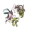
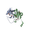
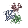
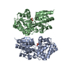
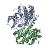
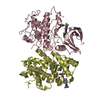
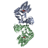
 Streptococcus pneumoniae (bacteria)
 X-RAY DIFFRACTION /
  Authors
 Citation
 Structure visualization
 Downloads & links
 Other downloads
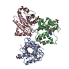

 PDBj
PDBj


 Assembly
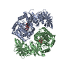
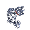
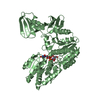

 Type: RNA linking / Mass: 404.161 Da / Num. of mol.: 2 / Source method: obtained synthetically / Formula: C9H14N2O12P2 / Comment: UDP*YM
 Type: D-saccharide, beta linking / Mass: 221.208 Da / Num. of mol.: 2
 Mass: 18.015 Da / Num. of mol.: 667 / Source method: isolated from a natural source / Formula: H2O
 Sample preparation
 / Beamline: BL17U / Wavelength: 0.97923 Å
 Processing