 Movie
Movie Controller

[English] 日本語
 Yorodumi
- PDB-4ppf: Mycobacterium tuberculosis RecA citrate bound low temperature str... -
+ Open data

- Basic information
| Entry | Database: PDB / ID: 4ppf | ||||||
|---|---|---|---|---|---|---|---|
| Title | Mycobacterium tuberculosis RecA citrate bound low temperature structure IIA-N | ||||||
 Components Components | Protein RecA, 1st part, 2nd part | ||||||
 Keywords Keywords | HYDROLASE / HOMOLOGOUS RECOMBINATION / DNA REPAIR / ATPASE / RECOMBINASE / DNA BINDING PROTEIN / PLOOP CONTAINING NTPASE FOLD / ATP BINDING / HYDROLYSIS | ||||||
| Function / homology |  Function and homology information Function and homology informationDNA strand invasion / intron homing / intein-mediated protein splicing / UV protection / DNA strand exchange activity / SOS response / recombinational repair / ATP-dependent DNA damage sensor activity / ATP-dependent activity, acting on DNA / DNA endonuclease activity ...DNA strand invasion / intron homing / intein-mediated protein splicing / UV protection / DNA strand exchange activity / SOS response / recombinational repair / ATP-dependent DNA damage sensor activity / ATP-dependent activity, acting on DNA / DNA endonuclease activity / manganese ion binding / single-stranded DNA binding / endonuclease activity / Hydrolases; Acting on ester bonds / damaged DNA binding / response to antibiotic / DNA repair / DNA damage response / magnesium ion binding / ATP hydrolysis activity / ATP binding / cytosol / cytoplasm Similarity search - Function | ||||||
| Biological species |   Mycobacterium tuberculosis (bacteria) Mycobacterium tuberculosis (bacteria) | ||||||
| Method |  X-RAY DIFFRACTION / SYNCHROTRON / MOLECULAR REPLACEMENT / Resolution: 2.3 Å X-RAY DIFFRACTION / SYNCHROTRON / MOLECULAR REPLACEMENT / Resolution: 2.3 Å | ||||||
 Authors Authors | Chandran, A.V. / Prabu, J.R. / Patil, N.K. / Muniyappa, K. / Vijayan, M. | ||||||
 Citation Citation | Journal: J.Biosci. / Year: 2015 Title: Structural studies on Mycobacterium tuberculosis RecA: Molecular plasticity and interspecies variability Authors: Chandran, A.V. / Prabu, J.R. / Nautiyal, A. / Patil, K.N. / Muniyappa, K. / Vijayan, M. #1: Journal: Acta Crystallogr.,Sect.D / Year: 2008Title: Functionally important movements in RecA molecules and filaments: studies involving mutation and environmental changes Authors: Prabu, J.R. / Manjunath, G.P. / Chandra, N.R. / Muniyappa, K. / Vijayan, M. #2: Journal: Nucleic Acids Res. / Year: 2006Title: Crystallographic identification of an ordered C-terminal domain and a second nucleotide-binding site in RecA: new insights into allostery Authors: Krishna, R. / Manjunath, G.P. / Kumar, P. / Surolia, A. / Chandra, N.R. / Muniyappa, K. / Vijayan, M. #3: Journal: J.Bacteriol. / Year: 2003Title: Crystal structures of Mycobacterium smegmatis RecA and its nucleotide complexes Authors: Datta, S. / Krishna, R. / Ganesh, N. / Chandra, N.R. / Muniyappa, K. / Vijayan, M. #4: Journal: Proteins / Year: 2003Title: Structural studies on MtRecA-nucleotide complexes: insights into DNA and nucleotide binding and the structural signature of NTP recognition Authors: Datta, S. / Ganesh, N. / Chandra, N.R. / Muniyappa, K. / Vijayan, M. #5: Journal: Nucleic Acids Res. / Year: 2000Title: Crystal structures of Mycobacterium tuberculosis RecA and its complex with ADP-AlF(4): implications for decreased ATPase activity and molecular aggregation Authors: Datta, S. / Prabu, M.M. / Vaze, M.B. / Ganesh, N. / Chandra, N.R. / Muniyappa, K. / Vijayan, M. | ||||||
| History |
|
- Structure visualization
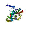
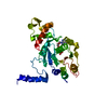
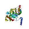
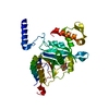
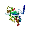
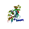
| Structure viewer | Molecule: MolmilJmol/JSmol |
|---|
- Downloads & links
-Download
| PDBx/mmCIF format | 4ppf.cif.gz | 77 KB | Display | PDBx/mmCIF format |
|---|---|---|---|---|
| PDB format | pdb4ppf.ent.gz | 55.6 KB | Display | PDB format |
| PDBx/mmJSON format | 4ppf.json.gz | Tree view | PDBx/mmJSON format | |
| Others |  Other downloads Other downloads |
-Validation report
| Arichive directory | https://data.pdbj.org/pub/pdb/validation_reports/pp/4ppfftp://data.pdbj.org/pub/pdb/validation_reports/pp/4ppf | HTTPS FTP |
|---|
-Related structure data
| Related structure data | 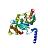 4oqfC 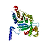 4po1C 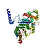 4po8C 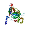 4po9C 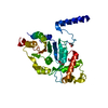 4poaC 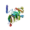 4ppgC  4ppnC 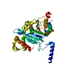 4ppqC 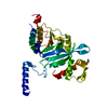 4pqfC  4pqrC 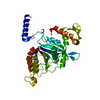 4pqyC 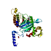 4pr0C 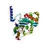 4psaC 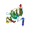 4pskC 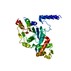 4psvC  4ptlC 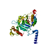 1g19S C: citing same article ( S: Starting model for refinement |
|---|---|
| Similar structure data |


-Links
 PDBj
PDBj


- Assembly
| Deposited unit | 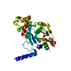
| ||||||||
|---|---|---|---|---|---|---|---|---|---|
| 1 |
| ||||||||
| Unit cell |
|
-Components
| #1: Protein | Mass: 37222.238 Da / Num. of mol.: 1 Source method: isolated from a genetically manipulated source Source: (gene. exp.) Mycobacterium tuberculosis (bacteria) / Gene: MT2806, MTV002.02c, recA, Rv2737c / Plasmid: PEJ135 / Production host: References: UniProt: P0A5U4, UniProt: P9WHJ3*PLUS, Hydrolases; Acting on ester bonds | ||||||
|---|---|---|---|---|---|---|---|
| #2: Chemical | ChemComp-FLC /   Mass: 189.100 Da / Num. of mol.: 1 / Source method: obtained synthetically / Formula: C6H5O7 Mass: 189.100 Da / Num. of mol.: 1 / Source method: obtained synthetically / Formula: C6H5O7 | ||||||
| #3: Chemical |   Mass: 22.990 Da / Num. of mol.: 2 / Source method: obtained synthetically / Formula: Na Mass: 22.990 Da / Num. of mol.: 2 / Source method: obtained synthetically / Formula: Na#4: Chemical | ChemComp-EDO /   Mass: 62.068 Da / Num. of mol.: 4 / Source method: obtained synthetically / Formula: C2H6O2 Mass: 62.068 Da / Num. of mol.: 4 / Source method: obtained synthetically / Formula: C2H6O2#5: Water | ChemComp-HOH / |  Mass: 18.015 Da / Num. of mol.: 110 / Source method: isolated from a natural source / Formula: H2O Mass: 18.015 Da / Num. of mol.: 110 / Source method: isolated from a natural source / Formula: H2OSequence details | THE PROTEIN IS EXPRESSED AS A 790 AMINO ACID RESIDUE PRECURSOR (UNIPROT P0A5U4). ONCE IT IS ...THE PROTEIN IS EXPRESSED AS A 790 AMINO ACID RESIDUE PRECURSOR (UNIPROT P0A5U4). ONCE IT IS RELEASED INTO THE CELL, THE CHAIN MTU RECA INTEIN (RESIDUES 252-691) IS CLEAVED OFF, CHAINS 1ST PART (RESIDUES 1-251) AND 2ND PART (RESIDUES 692-790) JOIN TOGETHER TO FORM THE MATURE PROTEIN. | |
-Experimental details
-Experiment
| Experiment | Method: X-RAY DIFFRACTION / Number of used crystals: 1 |
|---|
- Sample preparation
| Crystal | Density Matthews: 3.12 Å3/Da / Density % sol: 60.59 % |
|---|---|
| Crystal grow | Temperature: 298 K / Method: vapor diffusion, hanging drop / pH: 5.8 Details: 15% PEG 3350, 10% PEG 5000 MME, 0.2M AMMONIUM ACETATE, 0.1M SODIUM CITRATE, pH 5.8, VAPOR DIFFUSION, HANGING DROP, temperature 298K |
-Data collection
| Diffraction | Mean temperature: 100 K |
|---|---|
| Diffraction source | Source: SYNCHROTRON / Site: ESRF  / Beamline: BM14 / Wavelength: 0.976 Å / Beamline: BM14 / Wavelength: 0.976 Å |
| Detector | Type: MARMOSAIC 225 mm CCD / Detector: CCD / Date: Apr 27, 2013 / Details: Bent collimating mirror and toroid |
| Radiation | Monochromator: Si(111) monochromator / Protocol: SINGLE WAVELENGTH / Monochromatic (M) / Laue (L): M / Scattering type: x-ray |
| Radiation wavelength | Wavelength: 0.976 Å / Relative weight: 1 |
| Reflection | Resolution: 2.3→46.34 Å / Num. obs: 19963 / Redundancy: 10.5 % / Biso Wilson estimate: 41 Å2 / Rmerge(I) obs: 0.084 / Net I/σ(I): 18 |
| Reflection shell | Resolution: 2.3→2.42 Å / Redundancy: 6.7 % / Rmerge(I) obs: 0.876 / Mean I/σ(I) obs: 2 / Num. unique all: 2835 |
- Processing
| Software |
| ||||||||||||||||||||||||||||||||||||||||||||||||||||||||||||
|---|---|---|---|---|---|---|---|---|---|---|---|---|---|---|---|---|---|---|---|---|---|---|---|---|---|---|---|---|---|---|---|---|---|---|---|---|---|---|---|---|---|---|---|---|---|---|---|---|---|---|---|---|---|---|---|---|---|---|---|---|---|
| Refinement | Method to determine structure: MOLECULAR REPLACEMENT Starting model: PDB ENTRY 1G19 Resolution: 2.3→30 Å / Cor.coef. Fo:Fc: 0.96 / Cor.coef. Fo:Fc free: 0.937 / SU B: 6.043 / SU ML: 0.144 / Cross valid method: THROUGHOUT / ESU R: 0.239 / ESU R Free: 0.207 / Stereochemistry target values: MAXIMUM LIKELIHOOD / Details: HYDROGENS HAVE BEEN ADDED IN THE RIDING POSITIONS
| ||||||||||||||||||||||||||||||||||||||||||||||||||||||||||||
| Solvent computation | Ion probe radii: 0.8 Å / Shrinkage radii: 0.8 Å / VDW probe radii: 1.2 Å / Solvent model: MASK | ||||||||||||||||||||||||||||||||||||||||||||||||||||||||||||
| Displacement parameters | Biso mean: 47.901 Å2
| ||||||||||||||||||||||||||||||||||||||||||||||||||||||||||||
| Refinement step | Cycle: LAST / Resolution: 2.3→30 Å
| ||||||||||||||||||||||||||||||||||||||||||||||||||||||||||||
| Refine LS restraints |
| ||||||||||||||||||||||||||||||||||||||||||||||||||||||||||||
| LS refinement shell | Resolution: 2.3→2.36 Å / Total num. of bins used: 20
|
