Type: MARMOSAIC 225 mm CCD / Detector: CCD / Date: Nov 5, 2013
Radiation
Protocol: SINGLE WAVELENGTH / Monochromatic (M) / Laue (L): M / Scattering type: x-ray
Radiation wavelength
Wavelength: 1.03 Å / Relative weight: 1
Reflection
Resolution: 1.3→50 Å / Num. obs: 69082 / % possible obs: 94.4 % / Redundancy: 2.9 % / Rmerge(I) obs: 0.04 / Χ2: 1.048 / Net I/av σ(I): 20.845 / Net I/σ(I): 13.5 / Num. measured all: 200829
Reflection shell
Diffraction-ID: 1 / Rejects: _
Resolution (Å)
Redundancy (%)
Rmerge(I) obs
Num. unique all
Χ2
% possible all
1.3-1.32
3
0.561
3442
1.071
95.5
1.32-1.35
3
0.506
3467
1.075
96.6
1.35-1.37
2.9
0.436
3471
1.068
95.8
1.37-1.4
2.9
0.387
3474
1.063
95.5
1.4-1.43
2.9
0.332
3455
1.049
96
1.43-1.46
2.9
0.269
3439
1.032
95.3
1.46-1.5
2.8
0.22
3429
1.03
94.8
1.5-1.54
2.8
0.204
3440
1.077
94.8
1.54-1.59
2.7
0.161
3411
1.054
93.5
1.59-1.64
2.7
0.133
3372
1.082
92.9
1.64-1.7
2.7
0.119
3341
1.03
92.4
1.7-1.76
2.7
0.093
3329
1.078
91.6
1.76-1.84
2.8
0.075
3329
1.004
91.4
1.84-1.94
2.8
0.063
3300
1.057
90.2
1.94-2.06
2.8
0.057
3249
1.028
89.2
2.06-2.22
2.7
0.051
3339
1.017
90.6
2.22-2.45
2.8
0.043
3472
1.028
94
2.45-2.8
3.2
0.028
3671
1.105
99.2
2.8-3.53
3.5
0.02
3753
1.013
99.9
3.53-50
3.4
0.016
3899
1.005
99
-
Processing
Software
Name
Version
Classification
REFMAC
5.7.0032
refinement
HKL-2000
datareduction
PDB_EXTRACT
3.14
dataextraction
Refinement
Method to determine structure: SAD / Resolution: 1.304→41.95 Å / Cor.coef. Fo:Fc: 0.975 / Cor.coef. Fo:Fc free: 0.964 / SU B: 1.683 / SU ML: 0.031 / Cross valid method: THROUGHOUT / σ(F): 0 / ESU R: 0.046 / ESU R Free: 0.048 / Stereochemistry target values: MAXIMUM LIKELIHOOD Details: HYDROGENS HAVE BEEN ADDED IN THE RIDING POSITIONS U VALUES : REFINED INDIVIDUALLY
Rfactor
Num. reflection
% reflection
Selection details
Rfree
0.1743
1873
2.7 %
RANDOM
Rwork
0.1322
67014
-
-
obs
0.1333
68887
93.68 %
-
Solvent computation
Ion probe radii: 0.8 Å / Shrinkage radii: 0.8 Å / VDW probe radii: 1.2 Å / Solvent model: MASK
In the structure databanks used in Yorodumi, some data are registered as the other names, "COVID-19 virus" and "2019-nCoV". Here are the details of the virus and the list of structure data.
Jan 31, 2019. EMDB accession codes are about to change! (news from PDBe EMDB page)
EMDB accession codes are about to change! (news from PDBe EMDB page)
The allocation of 4 digits for EMDB accession codes will soon come to an end. Whilst these codes will remain in use, new EMDB accession codes will include an additional digit and will expand incrementally as the available range of codes is exhausted. The current 4-digit format prefixed with “EMD-” (i.e. EMD-XXXX) will advance to a 5-digit format (i.e. EMD-XXXXX), and so on. It is currently estimated that the 4-digit codes will be depleted around Spring 2019, at which point the 5-digit format will come into force.
The EM Navigator/Yorodumi systems omit the EMD- prefix.
Related info.:Q: What is EMD? / ID/Accession-code notation in Yorodumi/EM Navigator
Yorodumi is a browser for structure data from EMDB, PDB, SASBDB, etc.
This page is also the successor to EM Navigator detail page, and also detail information page/front-end page for Omokage search.
The word "yorodu" (or yorozu) is an old Japanese word meaning "ten thousand". "mi" (miru) is to see.
Related info.:EMDB / PDB / SASBDB / Comparison of 3 databanks / Yorodumi Search / Aug 31, 2016. New EM Navigator & Yorodumi / Yorodumi Papers / Jmol/JSmol / Function and homology information / Changes in new EM Navigator and Yorodumi
 Movie
Movie Controller

 Yorodumi
 Open data
 Basic information
 Components
 Keywords
 Function and homology information
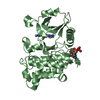
 Xanthomonas axonopodis pv. citri (bacteria)
 X-RAY DIFFRACTION /
  Authors
 Brazil, 1items
  Citation
 Structure visualization
 Downloads & links
 Other downloads


 PDBj
PDBj
 Assembly
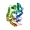

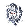
 Mass: 40.078 Da / Num. of mol.: 1 / Source method: obtained synthetically / Formula: Ca
 Mass: 18.015 Da / Num. of mol.: 323 / Source method: isolated from a natural source / Formula: H2O
 Sample preparation
 Processing